UNIGE researchers have discovered a new nano-structure that lies at the center of our cellular skeleton; this discovery will allow to better understand how the cell maintains its architecture as well as the pathologies associated with dysfunctions of this
Credit: © UNIGE
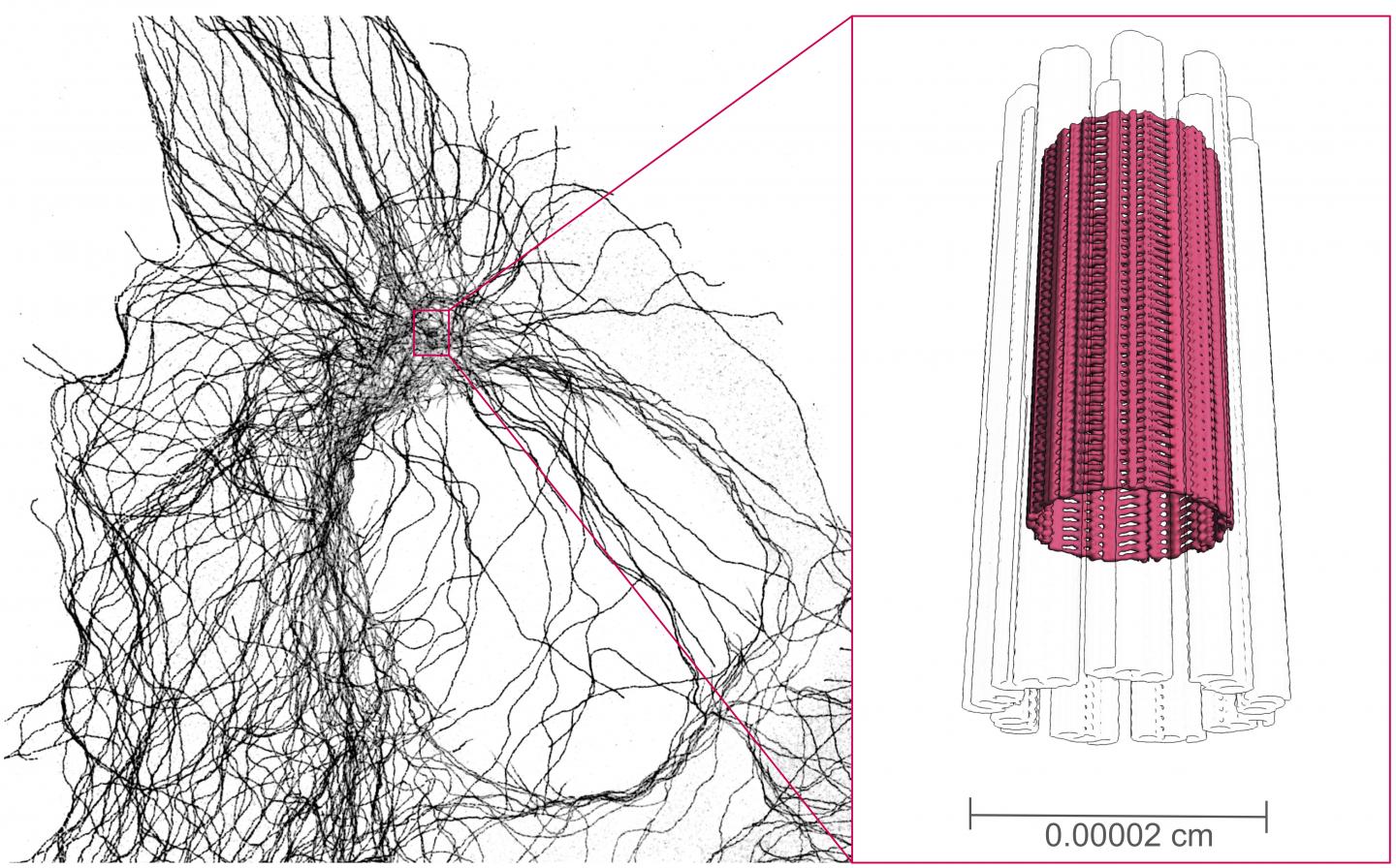
All animal cells have an organelle called a centrosome, which is essential to the organization of their cell skeleton. The centrosome plays fundamental roles, especially during cell division, where it allows equal sharing of genetic information between two daughter cells. When the cells stop dividing, the centrioles, cylindrical structures composed of microtubules at the base of the centrosome, migrate to the plasma membrane and allow the formation of primary and mobile cilia, which are used respectively for the transfer of information and the genesis of movement. While performing these crucial biological functions, centrioles are therefore subjected to many physical forces, which they must resist. Scientists from the University of Geneva (UNIGE) have discovered an internal structure at the center of these nano-cylinders, a real cellular scaffolding that maintains the physical integrity of this organelle. This study, published in the journal Science Advances, will provide a better understanding of the functions of the centriole and the pathologies associated with its dysfunction.
The centrioles, cylindrical nano-structures, form the centrosome, the main microtubule organizing center of the cell skeleton, and the cilia, real cellular antennas. Defects in the assembly or functioning of the centriole can lead to pathologies in humans, such as ciliopathies, retinal disorders that can cause loss of vision.
Super-powered microscopes
Centrioles, formed by microtubules, are components of the cell skeleton. “They have a canonical organization defined by nine triplets of microtubules that must be maintained as a structural unit in order to resist the various forces they face during their cellular functions,» explains Paul Guichard, Professor in the Department of Cell Biology of the Faculty of Science at UNIGE. The group of Paul Guichard and Virginie Hamel, a researcher at the Department of Cell Biology and co-leader of the study, discovered an internal scaffolding for this organelle using high-powered electron microscopes, in collaboration with researchers at the University of Basel and the Helmholtz Campus in Neuherberg, Germany. “This study allowed to analyze centrioles of four different species and to demonstrate that this inner scaffold is present systematically”, reports Maeva Le Guennec, a UNIGE researcher and first author of the study.
“We then investigated which centriolar proteins were located in this new structure”, says Virginie Hamel. To do this, the UNIGE researchers used an innovative super-resolution method, called expansion microscopy, which makes it possible to inflate cells without deforming them in order to observe their internal organization. Thus, they were able to identify four proteins that are located at the level of this inner scaffold.
Towards a better understanding of retinal degeneration
“We realized that the four proteins we identified are associated with pathologies related to retinal degeneration”, notes Virginie Hamel. The loss of retinal photoreceptors is possibly due to a failure to maintain the microtubule doublets present in these specialized cells. “We now intend to discover the possible link between such a structural maintenance defect and retinal disorders, in order to pave the way for a better understanding of this pathology”, concludes Paul Guichard.
###
Media Contact
Paul Guichard
[email protected]
41-223-796-750
Related Journal Article
http://dx.

