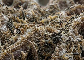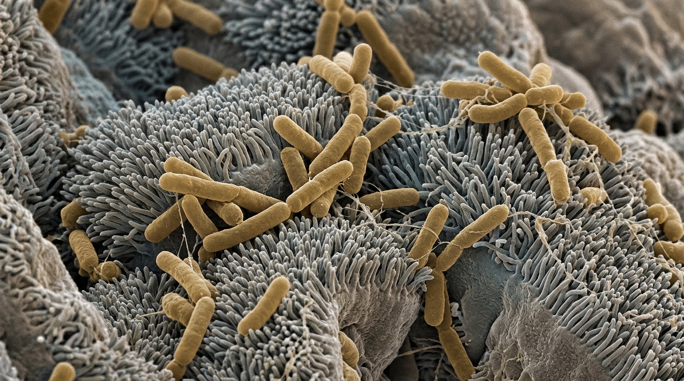
A comprehensive systematic review published in Frontiers in Cellular and Infection Microbiology demonstrates that the gut commensal bacterium Faecalibacterium actively counteracts metabolic dysfunction-associated fatty liver disease across multiple biological pathways. Metabolic dysfunction-associated fatty liver disease now represents the leading global cause of chronic liver disorders, with an estimated pooled prevalence of 38.77 percent, and can progress from simple steatosis to cirrhosis or hepatocellular carcinoma. By synthesizing preclinical mechanistic assays, animal models, and clinical investigations published through early 2026, researchers delineated how depletion of this beneficial anaerobic genus forms a hallmark of disease progression. While medical consensus has transitioned terminology from non-alcoholic fatty liver disease to metabolic dysfunction-associated fatty liver disease and steatotic liver disease, metabolic impairment remains central to pathogenesis. The synthesized evidence shows that Faecalibacterium mitigates liver injury through bioactive effectors, including short-chain fatty acids, microbial anti-inflammatory molecules, and extracellular vesicles, which systematically alleviate lipotoxicity, suppress inflammation, and restore mucosal defenses.
The relationship between intestinal ecology and hepatic pathology operates primarily through the bidirectional gut-liver axis, which channels gut-derived signals directly into hepatic tissue. Approximately 75 percent of portal vein blood supply originates in the intestine, exposing the liver to an extensive load of nutrients, antigens, and microbial toxins. Under normal physiological conditions, biliary transport delivers bile acids to the gut to modulate microbial balance and fortify epithelial integrity, providing feedback that maintains hepatic immune tolerance. However, gut dysbiosis disrupts this equilibrium, permitting toxic molecules like lipopolysaccharide to enter the portal bloodstream. In healthy individuals, the genus Faecalibacterium represents an abundant core taxon within the phylum Firmicutes, accounting for more than 5 percent of relative intestinal abundance. As strict anaerobes requiring dietary fibers, these bacteria depend on gut homeostasis. Observational clinical studies confirm that progressive depletion of Faecalibacterium directly correlates with disease severity as patients advance from simple steatosis to steatohepatitis and fibrosis.
Comparative genomics demonstrates that Faecalibacterium encompasses considerable diversity beyond the well-studied species Faecalibacterium prausnitzii, including Faecalibacterium duncaniae, Faecalibacterium hattorii, Faecalibacterium gallinarum, Faecalibacterium taiwanense, and Faecalibacterium langellae. Pan-genomic analyses establish that these species possess distinct carbohydrate-active enzyme repertoires, with glycoside hydrolase families particularly enriched in Faecalibacterium duncaniae and Faecalibacterium prausnitzii to ferment complex fibers into short-chain fatty acids. Functional traits also diverge between the two primary Faecalibacterium prausnitzii phylogroups. Phylogroup I strains, such as ATCC 27768 and M21/2, display elevated bile salt tolerance and preferentially utilize pectin and cellobiose while consuming acetate. Conversely, phylogroup II strains, including A2-165 and L2-15, exhibit high capacity for inulin utilization, generate abundant butyrate, produce specialized microbial anti-inflammatory molecules, secrete bioactive extracellular vesicles, and synthesize salicylic acid. Clinical observations link the depletion of phylogroup II strains directly with metabolic disorders and enteropathies, underscoring the clinical importance of species-level and strain-level functional variations.
The review collated quantitative findings from human clinical studies confirming the inverse relationship between Faecalibacterium abundance and metabolic hepatic injury. A subgroup analysis of a randomized controlled trial comprising 297 fatty liver patients showed that a Mediterranean lifestyle intervention elevated Faecalibacterium levels, which negatively correlated with hepatic steatosis and fibrosis-4 indices. In a prospective cohort study of 22 patients with metabolic dysfunction-associated fatty liver disease versus 44 healthy controls, bacterial depletion correlated with reduced fecal short-chain fatty acids; patients displayed elevated body mass index of 28.7 versus 20.8, alanine aminotransferase of 106.0 versus 15.5 units per liter, and higher fibrosis-4 scores. In metagenomic studies of 86 patients, Faecalibacterium was depleted in steatohepatitis cohorts with elevated aspartate aminotransferase of 72 versus 35 units per liter and glycated hemoglobin of 6.7 versus 6.0 percent. Additional trials demonstrated that dietary interventions restoring this taxon reduced total cholesterol and systemic inflammatory markers like C-reactive protein.
Mechanistically, Faecalibacterium directly interrupts the vicious cycle of insulin resistance and intracellular lipid accumulation driving hepatic steatosis. In metabolic liver disease, peripheral insulin resistance accelerates adipose lipolysis, causing an influx of free fatty acids into hepatocytes while hyperinsulinemia activates sterol regulatory element-binding protein 1c to stimulate de novo lipogenesis. Excess intracellular lipids activate protein kinase C, disrupting insulin receptor signaling and worsening insulin resistance. Faecalibacterium-derived short-chain fatty acids counter this cycle by stimulating intestinal G-protein-coupled receptors GPR41 and GPR43 to trigger glucagon-like peptide-1 secretion, which improves glycemic control, delays gastric emptying, and enhances satiety. Furthermore, short-chain fatty acids raise the cellular AMP-to-ATP ratio, activating AMP-activated protein kinase signaling. In peripheral tissues, this activation promotes glucose transporter type 4 translocation to facilitate glucose uptake. In hepatocytes, AMP-activated protein kinase inhibits sterol regulatory element-binding protein 1c to halt lipogenesis while activating the PGC-1alpha and PPARalpha axis to enhance fatty acid oxidation.
Beyond soluble short-chain fatty acids, Faecalibacterium secretes bacterial extracellular vesicles that act as nanoscale regulators of hepatic lipid metabolism and organelle integrity. Evidence indicates that these extracellular vesicles reprogram gut microbial metabolic activity, elevating levels of phosphatidylcholine. Because phosphatidylcholine is required for very low-density lipoprotein assembly and secretion, this restoration facilitates hepatic lipid export, preventing toxic triglyceride retention within hepatocytes. Extracellular vesicles also upregulate peroxisome proliferator-activated receptors and angiopoietin-like protein 4, augmenting mitochondrial fatty acid oxidation. Concurrently, these effectors alleviate hepatic organelle stress. In hepatocytes, excess lipids alter endoplasmic reticulum membranes and impair calcium transport by Sarco/endoplasmic reticulum calcium-ATPase pumps, triggering the unfolded protein response. Faecalibacterium extracellular vesicles restore membrane fluidity and pump function, while butyrate regulates chaperone protein GRP78 and downregulates pro-apoptotic CHOP transcription factor expression. This suppression blocks caspase-9 and caspase-3 activation, protecting hepatocytes from apoptosis and reducing damage-associated molecular pattern release.
Faecalibacterium provides robust protection against oxidative stress and non-apoptotic cell death by reinforcing cellular antioxidant defenses. Under persistent nutrient excess, mitochondrial electron transport chains become overloaded, causing reactive oxygen species overproduction that drives lipid peroxidation and depletes glutathione peroxidase 4, ultimately predisposing hepatocytes to ferroptosis. Acting as an epigenetic histone deacetylase inhibitor, butyrate promotes chromatin relaxation at the Nrf2 locus and accelerates Keap1-Nrf2 dissociation, driving Nrf2 nuclear translocation. Inside the nucleus, Nrf2 transcriptionally upregulates antioxidant enzymes, including heme oxygenase-1, glutathione S-transferases, and glutathione peroxidase 4, which detoxify lipid peroxides and inhibit ferroptotic death. Furthermore, butyrate directly inhibits NADPH oxidase activity, suppressing reactive oxygen species generation at the source. At the mitochondrial level, butyrate stimulates AMP-activated protein kinase to induce selective mitophagy, an essential quality-control mechanism that clears damaged mitochondria before they can release pro-apoptotic signals or trigger pro-inflammatory cascades.
To prevent simple steatosis from progressing to inflammatory steatohepatitis and fibrosis, Faecalibacterium mobilizes multi-targeted immunoregulatory mechanisms to suppress hepatic inflammation. When gut barrier disruption allows lipopolysaccharide to enter the portal bloodstream, Toll-like receptor 4 activation on Kupffer cells stimulates tumor necrosis factor-alpha and interleukin-1beta production. These cytokines activate I-kappa-B kinase beta, inducing I-kappa-B-alpha degradation and releasing nuclear factor-kappa-B to drive inflammatory gene transcription. Butyrate suppresses this pathway by inhibiting I-kappa-B kinase beta, increasing cyclic AMP to activate protein kinase A, and inhibiting histone deacetylase 3 to promote p65 acetylation, which reduces DNA binding. Additionally, Faecalibacterium produces microbial anti-inflammatory molecule, a specialized protein that directly blocks nuclear factor-kappa-B activation. In adaptive immunity, butyrate enhances regulatory T cell differentiation and interleukin-10 secretion, while oleic acid generated by specific strains suppresses inflammatory Th17 cells. Extracellular vesicles also promote anti-inflammatory M2b macrophage polarization by altering cellular respiration and cholesterol transport.
At the intestinal barrier, Faecalibacterium preserves mucosal integrity to prevent portal endotoxin translocation into the liver. Butyrate supplies up to 70 percent of the energy consumed by colonic epithelial cells, supporting cellular turnover and stimulating expression of critical tight junction proteins, including zonula occludens-1 and occludin. Concurrently, it stimulates goblet cells to secrete protective mucin-2, creating a robust dual-layer physical and biochemical defense. Crucially, research reveals that specific Faecalibacterium strains actively suppress mucin-degrading enzymes and mucosal degraders across the microbiome, preventing excessive mucus breakdown. Through metabolic cross-feeding, Faecalibacterium consumes acetate released by beneficial Bifidobacterium and Lactobacillus species while providing butyrate, fostering an optimized gut ecosystem. In animal models, administering strains such as F. prausnitzii EB-FPDK11 and UT1 repaired colonic architecture, elevated tight junction expression, and reduced circulating lipopolysaccharide levels, effectively blocking the gut-derived inflammatory signals that drive hepatic injury.
Despite compelling preclinical proof-of-concept findings, substantial biological and technological hurdles impede the clinical translation of Faecalibacterium as a live next-generation probiotic. Because these bacteria are strict anaerobes sensitive to ambient oxygen, maintaining cellular viability during large-scale fermentation, lyophilization, and storage remains challenging, although co-culturing with oxygen-scavenging Desulfovibrio or utilizing specialized lyoprotective formulations shows experimental promise. Furthermore, ensuring bacterial survival through gastric acidity and bile salts requires validated enteric delivery systems that currently lack clinical standardization. Industrial fermentation parameters remain unoptimized, human therapeutic dosing regimens are unestablished, and safety evaluations must address strain-specific antibiotic resistance, rare bacteremia risks in immunocompromised hosts, and potential immune overactivation. Consequently, researchers are emphasizing indirect strategies, including prebiotic fiber supplementation with inulin, fructooligosaccharides, or Euglena gracilis to boost endogenous strains, and postbiotic therapy using purified extracellular vesicles or calibrated butyrate doses, which demonstrate predictable dose-dependent efficacy without the technical hurdles of live bacterial administration.
Subject of Research: Mechanisms and therapeutic potential of Faecalibacterium in metabolic dysfunction-associated fatty liver disease
Article Title: Faecalibacterium: a promising gut commensal orchestrating metabolic, inflammatory, and barrier homeostasis in MAFLD
Article References: Wu, W., Xu, J., Cui, B., Guan, Y., Zhang, Q., & Liu, M. (2026). Faecalibacterium: a promising gut commensal orchestrating metabolic, inflammatory, and barrier homeostasis in MAFLD. Frontiers in Cellular and Infection Microbiology, 16, 1910351. https://doi.org/10.3389/fcimb.2026.1910351
Image Credits: AI Generated
DOI: 10.3389/fcimb.2026.1910351
Keywords: Faecalibacterium, Metabolic Dysfunction-Associated Fatty Liver Disease, Gut-Liver Axis, Short-Chain Fatty Acids, Butyrate, Microbial Anti-Inflammatory Molecule, Bacterial Extracellular Vesicles, Gut Microbiota Dysbiosis, Intestinal Barrier Integrity, Insulin Resistance, Hepatic Lipotoxicity, Endoplasmic Reticulum Stress, Ferroptosis, Next-Generation Probiotics, Prebiotics, Postbiotics, Macrophage Polarization, Tight Junction Proteins
News Source: Morgan Morrow. (November 17, 2026). Faecalibacterium Mitigates Metabolic Liver Disease Through Interconnected Multitargeted Pathways. Scienmag.