A comprehensive literature review published in Medicine Plus by researchers from Peking University People’s Hospital examines the current evidence for artificial intelligence and radiomics across the osteosarcoma imaging pathway, concluding that these technologies should function as clinical decision-support tools rather than autonomous replacements for radiologists or multidisciplinary teams. The researchers organized the evidence across core clinical tasks—radiographic triage, tumor segmentation, treatment response assessment, and risk prediction—while distinguishing osteosarcoma-specific studies from broader bone tumor investigations used as contextual evidence. Although algorithms demonstrate promise across multiple imaging modalities, the authors caution that high scores on conventional performance metrics such as the area under the receiver operating characteristic curve (AUC), Dice similarity coefficient, or concordance index do not guarantee that a model will perform reliably when deployed in another hospital or distinct patient population.
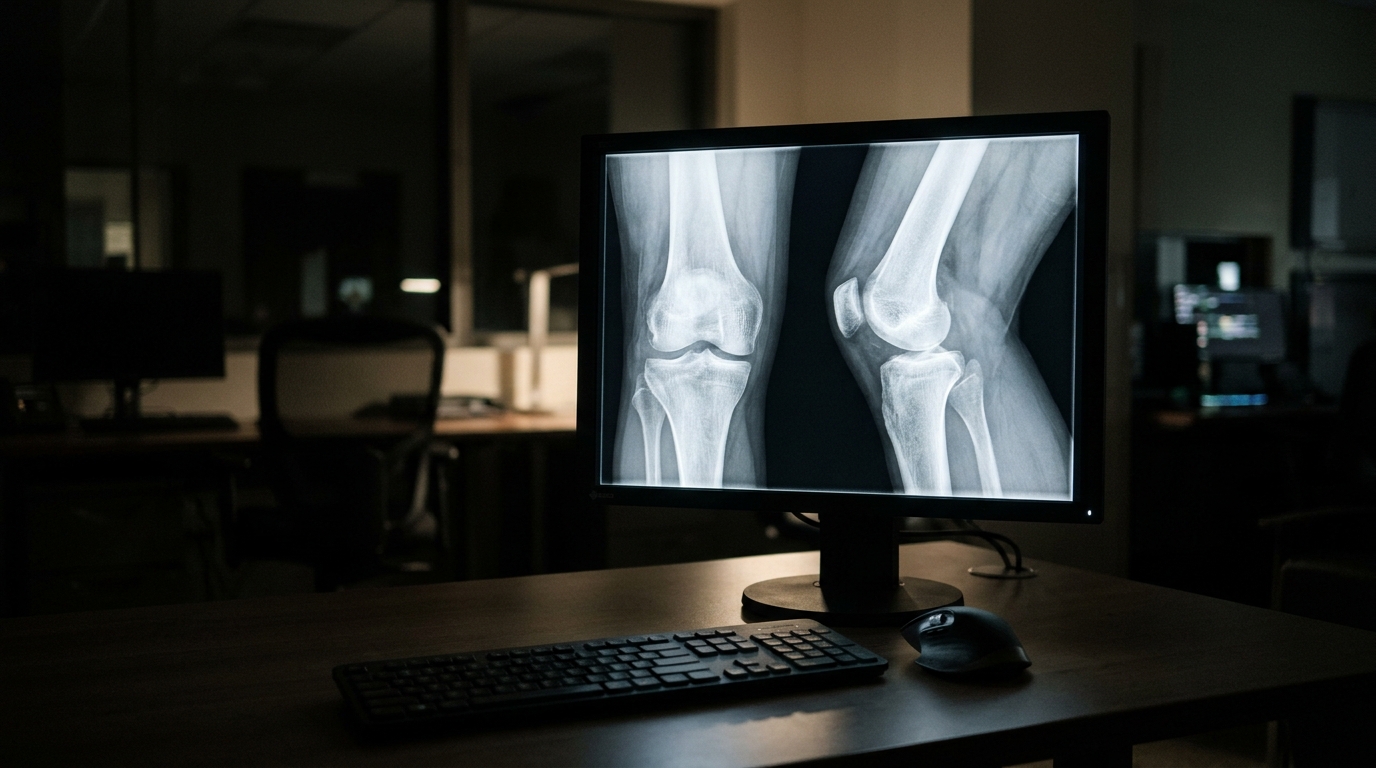
Among current applications, radiograph-based artificial intelligence appears closest to clinical workflow integration, with several models developed to detect and classify suspicious bone lesions on full-field radiographs to assist radiologists. Imaging plays a vital role throughout osteosarcoma care—including diagnosis, staging, treatment planning, response evaluation, and follow-up—where radiographs typically serve as the initial examination, complemented by computed tomography (CT) and magnetic resonance imaging (MRI) to assess local tumor extent, bone destruction, and pulmonary metastases. Because subtle early presentations, overlap with other bone diseases, and post-treatment alterations present major diagnostic challenges, the review suggests that the most realistic near-term clinical role for AI is supporting triage and referral decisions, such as prompting MRI scans, specialist referral, or biopsy planning, rather than rendering autonomous diagnoses.
Automated tumor segmentation on CT and MRI represents another active research domain designed to reduce labor-intensive manual contouring and enable consistent quantitative radiomic analysis. However, many osteosarcoma-specific segmentation studies remain constrained by relatively small cohorts of independent patients. The authors emphasize that evaluating thousands of individual image slices does not equate to analyzing thousands of independent clinical cases, noting that a favorable overall Dice score can mask significant local segmentation errors near critical joint surfaces, adjacent neurovascular structures, or small skip lesions. Furthermore, applying artificial intelligence to predict chemotherapy response, local recurrence, distant metastasis, and overall survival remains exploratory, as published studies vary widely in imaging timing, acquisition sequences, clinical endpoints, and validation strategies.
Moving machine learning models from retrospective developmental settings into routine clinical practice requires overcoming several interconnected translational challenges, starting with the fact that osteosarcoma is a rare and heterogeneous condition, making large, representative datasets difficult to assemble. Annotation and reference standards remain inconsistent across the field, while essential practices such as patient-level data partitioning, independent external validation, model calibration, subgroup analyses, and systematic failure-case reviews are not yet routinely reported. Additionally, multicenter translation must account for substantial variations in scanner hardware, imaging protocols, and clinical definitions. While explainability techniques can assist clinicians in inspecting model behaviors, the authors stress that they do not establish biological or clinical validity, underscoring the necessity of prospective trials, workflow safety testing, and ongoing performance monitoring to verify measurable clinical value.
Subject of Research: Artificial intelligence and radiomics in osteosarcoma imaging
Article Title: What will it take to bring AI into routine osteosarcoma imaging?
Article References: What will it take to bring AI into routine osteosarcoma imaging?. Original publication (Bibliographic details unavailable.)
Image Credits: AI Generated
DOI: Not provided
Keywords: osteosarcoma, artificial intelligence, radiomics, medical imaging, external validation, radiographic triage, tumor segmentation, clinical decision support, will, take, bring, routine
News Source: Nathaniel Bowman. (November 11, 2026). Review Outlines Evidence Needed to Bring AI into Routine Osteosarcoma Imaging. Scienmag.