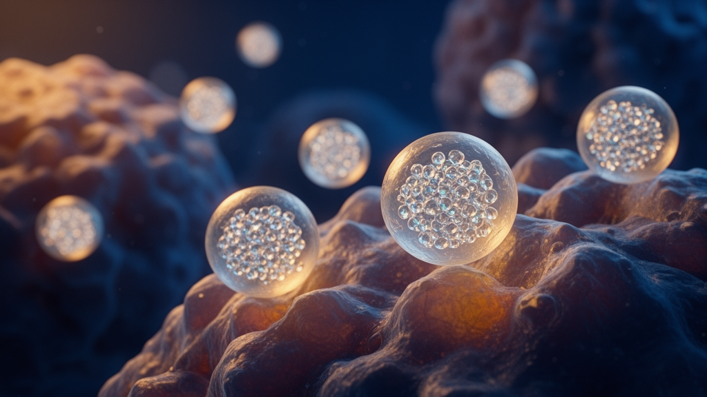
Researchers at Mansoura University in Egypt reported that a novel combination of melatonin and selenium nanoparticles significantly reduced tumor growth in a mouse model of cancer. The study, published in Cancer Nanotechnology in June 2026, evaluated the in vivo efficacy of melatonin-selenium nanoparticles (MSeNPs) in mice bearing Ehrlich ascites carcinoma (EAC). The findings indicate that the nanoparticle formulation outperformed individual treatments with melatonin, selenium, or selenium nanoparticles alone, suggesting a synergistic therapeutic effect. This approach aims to address the limitations of single-agent cancer therapies by combining agents with distinct biological properties into a single nanomedicine platform.
The research team, led by Hanaa M. Morad, A. F. Abdel-Aziz, and Mai M. Madkour, sought to investigate whether combining melatonin and selenium nanoparticles could enhance anticancer activity while improving safety profiles. Melatonin is a naturally occurring hormone known for its antioxidant and anti-angiogenic properties, while selenium is an essential trace element with established roles in immune function and cancer prevention. By encapsulating melatonin within selenium nanoparticles, the researchers created a formulation designed to improve drug delivery, stability, and bioavailability. The study utilized a mouse model of EAC, a widely used experimental system for evaluating anticancer agents, to assess the impact of MSeNPs on tumor progression and organ function.
The experimental design involved sixty-six female Swiss albino mice, which were divided into eleven groups of six animals each. The groups included negative controls, untreated EAC-bearing mice, and various treatment arms receiving sodium selenite, melatonin, selenium nanoparticles, or MSeNPs at doses of 5 and 10 mg/kg. The researchers tested both post-induction treatment and pretreatment protocols, where the substance was administered before tumor induction and continued afterward. This design allowed for a direct comparison of therapeutic versus preventive strategies and a detailed assessment of dose-dependent effects. All procedures were approved by the institutional ethics committee and followed the ARRIVE guidelines for reporting animal experiments.
Before in vivo testing, the researchers characterized the synthesized nanoparticles using scanning electron microscopy, transmission electron microscopy, and dynamic light scattering. The selenium nanoparticles exhibited a spherical shape with a mean hydrodynamic diameter of approximately 230 nm and a zeta potential of -30.5 mV, indicating good colloidal stability. The melatonin-selenium nanoparticles were larger, with an average size of 574.4 nm, and displayed a more negative zeta potential of -82.3 mV, suggesting enhanced stability. Fourier-transform infrared spectroscopy and X-ray diffraction confirmed the successful formation of the nanoparticles, revealing structural changes consistent with the interaction between melatonin and selenium. An acute toxicity study in mice showed no observable behavioral changes or mortality at doses up to 20 mg/kg, establishing a safety profile for the subsequent experiments.
The primary outcome of the study was the reduction in tumor volume, measured by the accumulation of ascitic fluid in the peritoneal cavity. Treatment with MSeNPs resulted in an 84% reduction in tumor volume compared to untreated controls. Both the 5 mg/kg and 10 mg/kg doses of MSeNPs produced significant reductions in tumor volume relative to treatments with selenium, selenium nanoparticles, or melatonin alone. Notably, the pretreatment group receiving MSeNPs at 10 mg/kg exhibited the lowest tumor volume, although this difference was not statistically significant compared to the post-treatment group at the same dose. The results suggest that the combination therapy exerts a potent cytotoxic effect on EAC cells, with the nanoparticle formulation offering superior efficacy over its individual components.
In addition to tumor suppression, the researchers evaluated the impact of MSeNPs on liver and kidney function, which are often compromised by cancer and its treatments. Serum levels of alanine aminotransferase (ALT) and aspartate aminotransferase (AST), markers of liver injury, were significantly elevated in untreated EAC mice. Treatment with MSeNPs, particularly at the 10 mg/kg dose, led to a marked decrease in these enzymes, indicating improved hepatic function. Similarly, serum urea and creatinine levels, which reflect renal function, were significantly reduced in the MSeNPs-treated groups compared to controls. The pretreatment groups generally showed better preservation of organ function than the treatment groups, highlighting the potential of MSeNPs to mitigate organ damage associated with tumor progression.
The study also investigated the anti-inflammatory and anti-angiogenic properties of the nanoparticles using real-time quantitative PCR and immunohistochemistry. MSeNPs treatment significantly downregulated the expression of tumor necrosis factor-alpha (TNF-α), a key pro-inflammatory cytokine, and vascular endothelial growth factor (VEGF), a critical driver of tumor angiogenesis. The reduction in VEGF expression was most pronounced in the pretreatment group, suggesting that early administration of MSeNPs may more effectively inhibit the formation of new blood vessels that support tumor growth. Immunohistochemical staining of tumor sections confirmed these molecular findings, showing reduced VEGF protein expression in treated mice. These results indicate that MSeNPs may disrupt the tumor microenvironment by suppressing both inflammation and angiogenesis.
Flow cytometry analysis revealed that MSeNPs shifted the mode of cell death from necrosis to apoptosis. In contrast, treatment with MSeNPs induced a significant increase in late apoptosis, with rates reaching approximately 38% in treated groups and 40.98% in the pretreatment group. This shift toward apoptotic cell death is considered a more favorable outcome in cancer therapy, as apoptosis is a controlled process that minimizes inflammatory damage to surrounding tissues. The nanoparticle formulation demonstrated superior pro-apoptotic activity compared to melatonin or selenium alone, suggesting that the combination enhances the ability to trigger programmed cell death in tumor cells.
Ultrastructural and histopathological examinations provided further evidence of the therapeutic effects of MSeNPs. Transmission electron microscopy of tumor cells showed morphological changes consistent with apoptosis, including nuclear shrinkage, cytoplasmic vacuolization, and mitochondrial swelling. In the liver, histological analysis revealed that MSeNPs treatment reduced the infiltration of EAC cells and restored hepatic architecture. Untreated mice exhibited severe liver damage, including vascular congestion, fibrosis, and hepatocyte necrosis. In contrast, mice treated with MSeNPs, particularly the pretreatment group, showed minimal pathological changes and near-normal liver histology. These findings support the conclusion that MSeNPs not only target the tumor but also protect healthy organs from the damaging effects of cancer.
The authors concluded that MSeNPs represent a promising nanotherapeutic strategy for cancer treatment, offering synergistic anticancer, anti-inflammatory, and anti-angiogenic effects. However, they noted that the study was limited to a single tumor model, which may restrict the generalizability of the findings to other types of cancer. The researchers emphasized the need for additional studies in different solid tumor models and human cancer cell lines to confirm the broader applicability of this combinatorial approach. Future investigations should also evaluate the long-term safety, pharmacokinetics, and bioaccumulation of selenium in the body to fully establish the translational potential of MSeNPs. Despite these limitations, the study provides preliminary evidence that melatonin-selenium nanoparticles could serve as an effective and safe option for both preventive and therapeutic cancer applications.
Beyond structural morphology, the investigation detailed the precise chemical preparation and physical stability of the nanoformulation. The synthesis of melatonin-selenium nanoparticles relied on reducing selenious acid and ascorbic acid in the presence of melatonin, yielding a crimson reaction product composed of 95 percent melatonin, 1 percent elemental selenium, and 4 percent ascorbic acid. Suspended in a carboxymethyl cellulose solution, the formulation achieved an encapsulation efficiency of approximately 69 percent and a drug loading capacity of 65.6 percent. Dynamic light scattering demonstrated a polydispersity index of 0.251 for the combined nanoparticles and 0.107 for selenium nanoparticles alone, indicating narrow particle size distributions and minimal aggregation. Furthermore, stability assessments conducted at both four degrees Celsius and room temperature revealed no significant alterations in hydrodynamic size, polydispersity index, or zeta potential over four weeks of storage.
Spectroscopic and crystallographic evaluations confirmed molecular bonding and crystalline reorganization within the hybrid system. Fourier-transform infrared spectroscopy revealed that characteristic bands of melatonin, notably carbonyl stretching at 1615.89 reciprocal centimeters and amine vibrations, were retained in the nanoparticle spectrum with reduced intensity, band broadening, and a shift of the carbonyl band to 1610.69 reciprocal centimeters. Increased infrared absorption between 600 and 800 reciprocal centimeters further corroborated the incorporation of elemental selenium. Complementary X-ray diffraction patterns verified that pure selenious acid exhibited sharp crystalline peaks at diffraction angles of 19.5 and 24.6 degrees, while melatonin showed semi-crystalline reflections between 16.5 and 26.1 degrees. In contrast, the melatonin-selenium nanoparticles displayed peak broadening alongside an entirely new diffraction peak at 10.8 degrees, demonstrating a distinct crystalline phase and a reduction in crystallite size arising from chemical interactions between both active compounds.
The study also utilized rigorous blinding and semi-quantitative ordinal scoring systems to grade histopathological damage in tumor tissue and hepatic structures. In carcinoma sections, untreated controls exhibited the lowest pathological score of two, characterized by dense mitotic figures and negligible necrosis or apoptosis. In contrast, tumor tissue from mice pretreated with melatonin-selenium nanoparticles at ten milligrams per kilogram attained the maximum pathological score of eight, marked by extensive apoptosis, ghost cell formation, and a complete absence of mitotic activity. Blinded assessment of liver specimens similarly documented substantial tissue protection under Cohen’s kappa evaluation. Untreated tumor-bearing mice registered a severe liver injury score of thirteen, defined by extensive portal fibrosis, hydropic degeneration, and vascular congestion. Mice receiving the ten-milligram-per-kilogram nanoparticle pretreatment exhibited a near-normal liver score of one, underscoring pronounced systemic preservation alongside localized tumor suppression.
Subject of Research: In vivo evaluation of melatonin-selenium nanoparticles in a mouse model of Ehrlich ascites carcinoma
Article Title: In vivo evaluation of the anticancer efficacy of melatonin–selenium nanoparticles in a mouse model
Article References: Morad, H. M., Abdel-Aziz, A. F., & Madkour, M. M. (2026). In vivo evaluation of the anticancer efficacy of melatonin–selenium nanoparticles in a mouse model. Cancer Nanotechnology, 17(1), Article 32. https://doi.org/10.1186/s12645-026-00389-8
Image Credits: AI Generated
DOI: 10.1186/s12645-026-00389-8
Keywords: melatonin, selenium nanoparticles, cancer nanotechnology, Ehrlich ascites carcinoma, anticancer therapy, apoptosis, angiogenesis, mouse model, drug delivery, synergistic therapy, vivo, evaluation
News Source: Nathaniel Bowman. (November 10, 2026). Melatonin-Selenium Nanoparticles Reduce Tumor Volume in Mice. Scienmag.