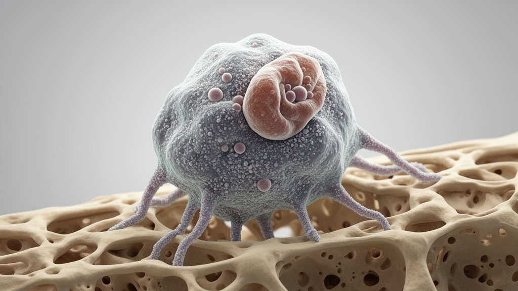
Researchers have identified macrophages as a central biological link between systemic aging and osteoporosis, utilizing a comprehensive integration of genetic and single-cell transcriptomic data. Published in Aging Cell, the study provides genetic evidence for a bidirectional causal relationship between these two conditions, moving beyond simple observational correlations. By combining large-scale genome-wide association studies (GWAS) with single-cell RNA sequencing (scRNA-seq) of bone marrow, the team uncovered specific molecular mechanisms that connect the deterioration of bone mass to the broader aging process. This integrative approach highlights the role of immune cells in skeletal health, suggesting that interventions targeting macrophage function could potentially address both age-related bone loss and systemic aging.
The study addressed a critical gap in understanding how local bone disorders interact with global aging. While it is well established that aging accelerates bone loss, the reverse relationship—where bone pathology influences systemic aging—had been less clearly defined. The researchers employed a multi-faceted analytical strategy to investigate this link. They utilized individual-level phenotypic data from the UK Biobank, specifically retaining 278,164 participants with complete information on heel bone mineral density (BMD), fracture status, and age. This large cohort allowed for robust statistical modeling of the relationship between skeletal health and aging metrics, providing a foundational observational basis for the subsequent genetic analyses.
To move from association to causation, the team applied Mendelian Randomization (MR) analyses using GWAS summary statistics. These genetic instruments allowed the researchers to infer causal directions between aging and osteoporosis traits. The results consistently supported a bidirectional causal relationship. Using inverse-variance weighted estimates, higher genetically determined GrimAge, a measure of biological aging, was associated with lower estimated BMD and higher fracture risk. Conversely, genetically determined fracture history was causally linked to higher GrimAge and lower mvAge, a multivariate anti-aging metric. These findings suggest that the molecular interplay between aging and bone health is not merely a one-way street, but a complex feedback loop where each condition influences the other.
Identifying the specific cell types driving this relationship required high-resolution cellular analysis. The researchers integrated two distinct single-cell transcriptomics datasets: one focusing on bone marrow niche organization and another on osteoclastogenesis. Because bone remodeling cells like osteoblasts and osteoclasts are relatively rare and technically challenging to capture, the team used deep learning-based methods, specifically scVI and scANVI, to integrate the data. This approach successfully merged the datasets, allowing for a comprehensive atlas of bone marrow cell populations. Validation confirmed that shared cell types, such as B cells and neutrophils, co-clustered effectively, while osteoclasts aligned with their progenitor monocytes, ensuring the biological integrity of the integrated atlas.
Single-cell GWAS (scGWAS) analysis was then performed to map GWAS signals to specific cell types. While mesenchymal stem cells and osteoblasts were linked to BMD, and osteoclasts to aging, macrophages emerged as the pivotal mediator connecting both processes. The analysis identified key genes within macrophage-specific modules, including transforming growth factor beta 1 (TGFB1) and cathepsin B (CTSB). TGFB1 is a known regulator of macrophage polarization, while CTSB is involved in osteoclastogenesis. The presence of these genes in modules associated with both aging and osteoporosis supported the hypothesis that macrophages act as a communication bridge between systemic aging and local bone remodeling.
To further characterize the age-related changes in macrophages, the team analyzed two independent mouse bone marrow datasets spanning different ages. Both datasets showed a consistent and substantial decline in the proportion of bone marrow macrophages with advancing age. Re-clustering identified five distinct macrophage subpopulations: IFN-stimulated, proliferating, MHC-II+, lipid-associated, and inflammatory. Compositional analysis revealed that the proportion of lipid-associated macrophages increased with age, while IFN-stimulated and inflammatory subsets decreased. Notably, MHC-II+ macrophages increased during late life and were specifically associated with both eBMD and mvAge in the scGWAS analysis, suggesting a specialized role for this subset in bone aging.
Single-cell weighted gene co-expression network analysis (scWGCNA) was used to identify gene networks associated with macrophage abundance during aging. The analysis identified a specific gene module, labeled “Gainsboro,” consisting of 171 genes that showed the strongest positive correlation with macrophage proportion and negative correlation with age. Gene Ontology enrichment analysis indicated that these genes were predominantly involved in epigenetic regulatory processes, including chromatin remodeling and transcriptional regulation. Genes such as BRD2 and BAZ2A, associated with histone acetylation, were highlighted, suggesting that age-related changes in macrophages are linked to alterations in epigenetic mechanisms within the bone marrow microenvironment.
Shared genetic methods were employed to identify novel genes involved in both aging and osteoporosis. Conjunctional false discovery rate (conjFDR) analysis identified approximately 160 SNPs significantly associated with both eBMD and mvAge. eQTL mapping linked these SNPs to 43 genes, with HNRNPUL1 (Heterogeneous Nuclear Ribonucleoprotein U Like 1) being the only gene overlapping with the “Gainsboro” module identified by scWGCNA. HNRNPUL1 is located in close physical proximity to TGFB1 on the chromosome. Mendelian Randomization analysis provided evidence for a causal relationship between HNRNPUL1 expression and aging-related phenotypes, while colocalization analysis confirmed that this association is mediated through the regulation of HNRNPUL1 gene expression. These results suggest that HNRNPUL1 is a novel genetic factor influencing both aging and bone health.
The functional relationship between HNRNPUL1 and TGFB1 was further explored through trajectory analysis of osteoclast differentiation. The expression levels of both genes decreased during macrophage differentiation into osteoclasts, suggesting they may regulate macrophage fate decisions. Although no significant correlation was found with classical M1/M2 polarization states, both genes showed strong associations with “Proliferating” and “MHC-II+” macrophage subclusters. Virtual knockout analyses indicated that neither gene directly regulates the expression of the other. However, computational predictions suggested a high potential for HNRNPUL1, an RNA-binding protein, to bind TGFB1 mRNA. This implies an HNRNPUL1-TGFB1 axis that may regulate macrophage function and bone aging through post-transcriptional mechanisms, such as RNA splicing.
The study acknowledges several limitations that temper the interpretation of these findings. The integration of GWAS summary statistics with single-cell data relies on average expression within cell populations, which may obscure individual cell-level variations. The scarcity of publicly available single-cell eQTL data for bone tissue limited the depth of the analysis. Additionally, the limited number of macrophages captured in the datasets constrained the resolution of subclustering, potentially affecting the characterization of rare macrophage subsets. The functional role of the HNRNPUL1-TGFB1 axis remains to be validated through experimental studies. Despite these limitations, the study provides a robust genetic and cellular framework for understanding the link between aging and osteoporosis.
These findings offer new perspectives on the pathogenesis of age-associated bone loss. By identifying macrophages as key mediators and highlighting the role of TGFB1 and HNRNPUL1, the research points to potential therapeutic targets for delaying systemic aging and preventing osteoporosis. This integrative approach underscores the importance of considering the immune system in skeletal biology and opens new avenues for research into the molecular mechanisms of bone aging.
The study utilized a diverse array of genomic resources to establish the genetic architecture of bone aging. For osteoporosis traits, the researchers employed summary statistics from the GEFOS Consortium, including datasets for estimated bone mineral density (eBMD) comprising 426,824 European samples, and fracture risk involving 53,184 cases and 373,611 controls. To represent systemic aging, the team integrated data from the GrimAge methylation clock, which included 34,710 European participants, and the mvAge multivariate metric. The mvAge variable was particularly notable for its large effective sample size of approximately 1.9 million participants, combining healthspan, lifespan, extreme longevity, frailty, and epigenetic aging to serve as a measure of senescence resistance. This extensive genetic foundation allowed for robust Mendelian Randomization analyses, where instrumental variables were selected to ensure strong exposure associations, with all F-statistics exceeding 10 to minimize weak instrument bias.
Observational data from the UK Biobank provided the initial phenotypic context for these genetic findings. After excluding outliers with heel bone mineral density values exceeding three standard deviations from the mean, the analysis of 278,164 participants revealed distinct sex-specific patterns in bone health. Generalized additive models indicated that while heel BMD declines with age in both sexes, the rate of this decline accelerates specifically in females after age 50. Furthermore, Kaplan–Meier survival analyses demonstrated that individuals with a history of fractures or lower heel BMD exhibited reduced survival rates. These observational trends supported the subsequent genetic inference of a bidirectional relationship, where aging influences bone density and bone pathology, in turn, impacts systemic aging metrics.
The identification of HNRNPUL1 as a key regulatory gene was supported by multiple layers of statistical validation. Beyond its overlap with the Gainsboro gene module, HNRNPUL1 was linked to 43 genes through expression quantitative trait locus (eQTL) mapping. The specific SNP rs12975920, located within the HNRNPUL1 gene body, showed significant associations with eBMD. Colocalization analysis further confirmed that the association between HNRNPUL1 expression and aging was mediated through shared causal variants, with a posterior probability of shared association signals exceeding 0.8. This convergence of genetic, transcriptomic, and functional evidence strengthens the case for HNRNPUL1 as a novel target in the HNRNPUL1-TGFB1 signaling axis, although the authors emphasize that experimental validation of this axis remains a necessary next step to confirm its functional role in bone aging.
Subject of Research: Genetic and single-cell analysis of the link between aging and osteoporosis
Article Title: Integrative Genetic and Single‐Cell Analysis Reveals Macrophages as Key Mediators Linking Aging and Osteoporosis
Article References: Li, X., Yang, M.-Y., Gai, S.-R., Wei, P., Gu, Z.-H., Han, M.-Y., Wu, Y.-Z., Yu, J.-S., Chen, W.-J., Liao, Z.-R., Gu, J.-X., Zhong, J.-D., Zhao, P.-P., Zhu, K., Cheung, C.-L., Karasik, D., & Zheng, H.-F. (2026). Integrative Genetic and Single‐Cell Analysis Reveals Macrophages as Key Mediators Linking Aging and Osteoporosis. Aging Cell, 25(11), Article e70812. https://doi.org/10.1111/acel.70812
Image Credits: AI Generated
DOI: 10.1111/acel.70812
Keywords: osteoporosis, aging, macrophages, genomics, single-cell RNA sequencing, Mendelian Randomization, bone density, TGFB1, HNRNPUL1, epigenetics, Integrative, Genetic
News Source: Juliet Wilcox. (November 9, 2026). Macrophages Link Aging and Osteoporosis Through Shared Genetic Pathways. Scienmag.