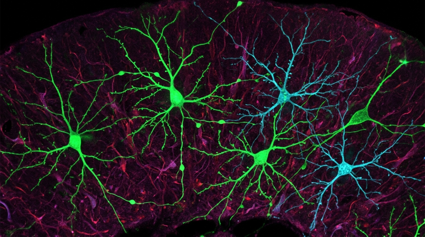
Chronic inhalation of formaldehyde vapour, the airborne component of the formalin fixative used in anatomy dissection halls, produced measurable neurodegeneration and neuroinflammation in the brains of adult male Wistar rats, according to a study published in BMC Neuroscience. After 28 days of exposure, the animals showed a marked reduction in the number and soma size of parvalbumin-expressing interneurons across the cerebellar Purkinje layer, the hippocampal CA1 region and the cerebral cortex, together with pronounced astrocytic and microglial activation in those regions and in the olfactory bulb.
Parvalbumin interneurons are fast-spiking inhibitory cells that regulate the timing and synchrony of network activity in the cortex and hippocampus. Their loss or dysfunction is a feature of several neurological conditions, which is why their depletion under formaldehyde exposure attracted the researchers’ attention. The study’s authors note that while the respiratory and mucosal effects of formaldehyde inhalation are well documented, the impact on the brain’s cellular architecture under a chronic, inhalation-based exposure paradigm had remained poorly characterised.
The exposure model was deliberately designed to exceed typical modern occupational breathing-zone concentrations, modelling a high-exposure, worst-case scenario relevant to poorly ventilated or historically under-regulated dissection settings. Formalin is the principal fixative used in gross anatomy dissection, and its vapour constitutes a recognised occupational hazard for anatomists, technicians and students during prolonged cadaveric exposure. The researchers describe their paradigm as closely mirroring occupational exposure conditions in anatomy dissection settings, though the concentration used sits above what is typical in contemporary laboratories.
Physically, the exposed animals fared poorly. They showed a significant, time-dependent decline in body weight gain, a reduction in absolute brain weight, and an elevation in brain formaldehyde concentration relative to controls. These systemic and tissue-level changes indicate that the inhaled vapour was absorbed and reached the central nervous system over the four-week exposure period.
Neurobehavioural testing reinforced the anatomical findings. Formaldehyde-exposed rats displayed impaired motor coordination, deficits in spatial memory and reduced exploratory behaviour. The regional distribution of the cellular damage offers a plausible correspondence with these functional deficits: Purkinje cell layer changes in the cerebellum align with motor coordination impairment, while hippocampal CA1 involvement corresponds with the spatial memory deficits observed in behavioural testing.
At the biochemical level, the exposed brains showed significantly elevated lipid peroxidation, a marker of oxidative damage to cell membranes, alongside depleted antioxidant enzyme activities. This shift in the balance between oxidant production and antioxidant defence provides a mechanistic context for the neuronal loss and glial activation seen under the microscope, although the study reports these changes as correlated observations within the same exposure paradigm rather than as a demonstrated causal sequence.
The quantitative immunohistochemical and morphometric analysis was notable for its breadth. The reduction in parvalbumin-positive interneuron number and soma size, and the accompanying activation of astrocytes and microglia, the brain’s resident immune and support cells, appeared in every region and layer examined rather than in a subset of regions. The authors state that this widespread pattern indicates that chronic formaldehyde inhalation produces measurable, widespread neurodegeneration and neuroinflammation in the rodent model. Astrocytic and microglial activation is interpreted in the study as a neuroinflammatory response accompanying the neuronal changes.
The findings carry practical weight for anatomical education and laboratory occupational health. The authors argue that the results underscore the need for stricter formaldehyde exposure controls in anatomical education and raise concern regarding the long-term neurological safety of personnel routinely exposed to formalin vapour in dissection halls. Because the exposure paradigm was designed as a worst-case scenario exceeding typical modern breathing-zone concentrations, the results speak most directly to poorly ventilated or historically under-regulated environments rather than to facilities operating at current occupational limits.
Several limitations frame the interpretation. Only male rats were examined, so the generalizability of the findings to female subjects remains to be established. The study was conducted in a rodent model, and the authors present it as modelling occupational exposure conditions rather than as a direct measurement of effects in humans. The article was published as an open access paper on 30 October 2026 and was shared early to provide faster access to peer-reviewed, accepted research; this version is subject to further edits and will be replaced by the final Version of Record.
For the anatomy departments, technical staff and students who spend prolonged hours over cadaveric tanks, the study adds a neurological dimension to a hazard already recognised for its respiratory and mucosal effects. The evidence base remains rodent-based and confined to a single sex, but the combination of behavioural deficits, oxidative stress markers, interneuron depletion and glial activation across every examined brain region gives the occupational health question a new and more specific focus: not only whether formaldehyde vapour irritates airways, but what it does to the cellular architecture of the brain under chronic, high-level inhalation exposure.
Subject of Research: Effects of chronic formaldehyde inhalation on interneurons and glial cells in the rat brain
Article Title: Chronic formaldehyde inhalation reduces parvalbumin immunoreactivity and modulates glial activation in the rat brain
Article References: Usende, I. L., Muhammad, B. A., Idris, N. A., Mobolaji, A. A., Shawulu, J. C., Oyelowo-Abdulraheem, F., Usende, E. U., Ja’afar, S., & Edem, E. E. (2026). Chronic formaldehyde inhalation reduces parvalbumin immunoreactivity and modulates glial activation in the rat brain. BMC Neuroscience. https://doi.org/10.1186/s12868-026-01064-9
Image Credits: AI Generated
DOI: 10.1186/s12868-026-01064-9
Keywords: formaldehyde, formalin, neurotoxicology, parvalbumin interneurons, neuroinflammation, oxidative stress, anatomy dissection, occupational health, Wistar rats, hippocampus, cerebellum, BMC Neuroscience
News Source: Cassandra Pierce. (November 6, 2026). Chronic formaldehyde inhalation damages key interneurons in rat brain study. Scienmag.