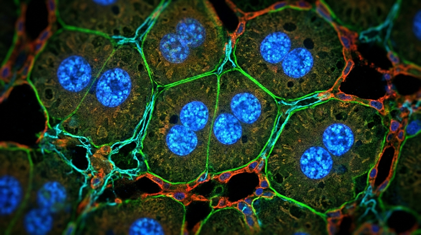
When bile cannot drain from the liver, the organ faces a silent crisis. Blocked bile ducts trigger inflammation, activate scar-forming cells and, if obstruction persists, push the tissue toward fibrosis and cirrhosis. Yet the liver also mounts an adaptive countermeasure: hepatocytes increase their DNA content, becoming polyploid, a shift that appears early after bile duct obstruction. A new study in rats now pinpoints which molecular switch drives that shift. Blocking the p38 MAPK signaling pathway sharply reduced the rise of high-ploidy hepatocytes after common bile duct ligation, while blocking the related JNK1/2 pathway changed nothing at all.
The work, published in the Egyptian Liver Journal, was carried out by Salome Kiparoidze, Ekaterine Bakuradze, Irina Modebadze, Levan Rusishvili and Diana Dzidziguri at Ivane Javakhishvili Tbilisi State University. The team set out to determine whether two well-characterized mitogen-activated protein kinase pathways, JNK1/2 and p38, participate in initiating polyploidization as an adaptive response in the cholestatic liver. Both pathways are known to be activated in hepatocytes during cholestasis, but their role in launching the polyploidization process had not been fully elucidated.
Cholestasis-induced liver injury is a major contributor to hepatic pathologies. Ligation of the common bile duct induces proliferation of epithelial cells and cholangiocytes, activates portal fibroblasts and hepatic myofibroblasts, and thereby promotes inflammation, fibrotic scar formation and ultimately fibrosis and cirrhosis. In conditions of mechanical obstruction, for example by gallstones or tumors, as well as in cirrhosis from primary biliary cholangitis or primary sclerosing cholangitis, liver transplantation can ultimately become necessary. Against this destructive backdrop, the liver under cholestatic conditions also shows adaptive responses to the increased functional load, and one of these is hepatocyte polyploidization, an increase in the number of high-ploidy cells.
The researchers had previously shown that during cholestatic conditions the liver responds to increased functional load without losing tissue mass by increasing the number of polyploid cells in the 4 C, 4 C×2 and 8 C classes, and that initiation of hepatocyte polyploidization proceeds through the inflammatory mediator TPL2 via the Tpl2–MEK–ERK signaling pathway. Because JNK1/2 and p38 are also activated in hepatocytes under cholestasis, the question was whether these stress-activated pathways also feed into the polyploidization program. To find out, the team ran experiments on adult white rats weighing 130 to 150 grams, using a cholestatic model created by common bile duct ligation, known as CBDL.
The study was conducted in two stages, each with three groups of ten animals. In the first stage, the JNK1/2 pathway was inhibited: one group of intact animals served as controls, a second group underwent CBDL with liver tissue collected 48 hours after surgery, and a third group received an intraperitoneal injection of the JNK inhibitor SP600125 at a dose of 10 milligrams per kilogram 20 hours after CBDL, with tissue also collected at 48 hours. The second stage mirrored this design with the p38 inhibitor SB203580 at the same dose and timing. The 48-hour endpoint was chosen because previous findings had shown that at this point the number of high-ploidy cells and the mitotic activity of liver parenchymal cells increase sharply, capturing polyploidization at its peak. Inhibitors were given 20 hours after CBDL, roughly four hours before hepatocytes would re-enter a second cell cycle, since hepatocytes need approximately 24 hours to complete one full cell cycle during the early regenerative response.
Proliferative activity was measured with the colchicine mitotic index, in which colchicine arrests dividing cells in metaphase so mitoses can be counted; for each sample 5,000 cells were analyzed and mitotic cells were expressed per 1,000 cells. Alterations in hepatocyte ploidy were assessed by Feulgen staining, which binds DNA, followed by quantitative image analysis with ImageJ software, measuring 300 cells per sample across 60 samples in total. Histology used hematoxylin and eosin staining of paraffin sections. All histological evaluation, mitotic index calculations and DNA quantification were performed in a blinded manner using coded slides. Data were analyzed with GraphPad Prism, with significance set at p less than 0.05, using Brown-Forsythe and Welch ANOVA with Dunnett’s T3 comparisons for mitotic activity and two-way repeated measures ANOVA with Tukey’s post-hoc tests for polyploidization.
The first series of experiments delivered a clear negative result for JNK. Forty-eight hours after CBDL, hepatocyte mitotic activity rose significantly compared with controls, reaching 24 per thousand. In animals that also received the JNK1/2 inhibitor, the number of mitotic cells at 48 hours did not change compared with the CBDL-only group. Histological images confirmed tissue alterations characteristic of cholestatic liver, including partial disruption of hepatic cord arrangement and narrowing of capillaries, and showed that the increased mitotic activity induced by CBDL was not altered by the inhibitor. The ploidy analysis told the same story: in CBDL animals the number of diploid cells fell approximately twofold while octaploid hepatocytes increased threefold, and the JNK inhibitor left the quantitative increase in octaploid cells unchanged. The authors concluded that JNK1/2 is not involved in the initiation of polyploidization at the early stage of cholestasis.
The second series pointed firmly at p38. As before, mitotic activity increased significantly 48 hours after CBDL, and the p38 inhibitor did not change the number of mitotic cells, so the pathway is not required for the proliferative burst itself. But the ploidy distribution diverged dramatically. Compared with controls, CBDL animals showed an increase in high-ploidy cells attributable to octaploid mononuclear and binuclear hepatocytes. Administration of SB203580 reduced the number of high-ploidy hepatocytes compared with the CBDL-only group, with statistically significant decreases in the proportions of 8 C and 4 C×2 cells at p less than 0.05. In other words, p38 inhibition significantly reduced the high-ploidy hepatocytes induced by CBDL without affecting mitotic activity, exactly the signature expected of a pathway governing the polyploidization program rather than ordinary cell division.
The authors propose a mechanistic logic for this division of labor. JNK is activated by inflammatory and stress-related signals including TNF and IL-1, reactive oxygen species, endoplasmic reticulum stress, toxins and pathogens, and its activation stimulates the AP-1 transcription factor, driving cellular responses such as cell death and fibrogenic signaling. Meanwhile, numerous signaling molecules activate p38 alongside JNK: MKK3 and MKK6 are the main MAP kinase kinases responsible for p38 activation, and MKK4, a known JNK activator, can also participate in p38 activation under certain conditions. On this basis the team hypothesized that under cholestasis, the initiation of polyploidization in response to functional load may depend on the p38 pathway operating in crosstalk with JNK, and the second series of results supports that hypothesis.
The conclusion, stated for the initial stage of cholestatic liver in adult white rats, is that under conditions of increased functional load caused by common bile duct ligation, the increase in the number of high-ploidy cells depends on activation of the p38 MAPK cascade, suggesting a crucial role for this pathway in managing the adaptive response. The findings carry the usual caveats of an animal model: the results come from 60 rats followed to a single 48-hour endpoint, and the authors note that the signaling pathways responsible for the cascade activation of hepatocyte polyploidization under cholestasis have not yet been fully elucidated. Even so, the study cleanly separates two stress-activated kinases that operate side by side in the injured liver, showing that at the earliest phase of obstructive cholestasis the adaptive expansion of polyploid hepatocytes runs through p38, while JNK1/2 stands apart from that particular program.
Subject of Research: Signaling pathways controlling the initiation of hepatocyte polyploidization in early cholestatic liver injury
Article Title: Role of JNK and p38 MAPK signaling pathways in the initiation of polyploidization in cholestatic liver
Article References: Kiparoidze, S., Bakuradze, E., Modebadze, I., Rusishvili, L., & Dzidziguri, D. (2026). Role of JNK and p38 MAPK signaling pathways in the initiation of polyploidization in cholestatic liver. Egyptian Liver Journal, 16(1), Article 61. https://doi.org/10.1186/s43066-026-00547-y
Image Credits: AI Generated
DOI: 10.1186/s43066-026-00547-y
Keywords: cholestasis, p38 MAPK, JNK1/2, polyploidization, common bile duct ligation, hepatocytes, liver fibrosis, SP600125, SB203580, rat model, ploidy, adaptive response
News Source: Ophelia Keating. (November 6, 2026). p38 MAPK Drives Early Polyploidization in Cholestatic Rat Liver, JNK Does Not. Scienmag.