Cerebrospinal fluid cytology remains a cornerstone of diagnostic evaluation in infectious, autoimmune, haemorrhagic and neoplastic disorders of the central nervous system, even as neuroimaging, molecular diagnostics and digital technologies advance around it. That is the central conclusion of a narrative review published in Acta Neuropathologica by a large expert group writing on behalf of the German Society for Cerebrospinal Fluid Diagnostics and Clinical Neurochemistry. The authors, led by Sandra Nay and Thomas Skripuletz, argue that the discipline’s value lies not in resisting new tools but in defining precisely where manual expertise must remain the reference standard and where automation can be safely introduced.
The review’s practical recommendations are unusually direct. The authors call for rapid cerebrospinal fluid processing, routine preparation of cytospin-based stained slides with expert microscopic review, and implementation of automated or digital methods only within clearly validated workflows. They describe manual chamber counting, particularly in the normal and low-cell-count range, as the gold standard for quantitative cell assessment, while expert microscopic examination remains the reference standard for qualitative cytomorphological assessment. Automated cell counters are increasingly implemented in routine laboratories and offer advantages where technical staffing is limited, but current systems show limited precision in samples with low and slightly elevated cell counts and limited morphological evaluation.
Pre-analytical handling emerges as a critical determinant of diagnostic accuracy. Cerebrospinal fluid has a low protein content and weak buffering capacity, making it highly susceptible to degradation. After sampling, the fluid’s pH rises from about 7.3 toward alkaline values within a few hours, promoting cellular autolysis, and these processes accelerate with higher ambient temperatures. Without stabilisation, leukocyte loss can be rapid: delays may result in significant reduction of lymphocytes ranging from minus 10 to minus 53 percent within five hours and up to minus 60 percent within 24 hours, as reported for lumbar and ventricular samples in neonatal and adult patients. Processing within one to two hours is therefore considered critical, and neutrophils and tumour cells are regarded as even more vulnerable to degradation.
The quantitative limits of automation are laid out in detail. A reliable lower limit of quantification for cerebrospinal fluid leukocytes on automated haematology analysers is still around 20 cells per microlitre, device dependent, with older systems possibly limited to around 50 cells per microlitre. Expert appraisal concludes that cell counts below 30 cells per microlitre require mandatory manual confirmation by chamber counting. A systematic review and meta-analysis by Waldrop and colleagues found good overall agreement between automated analysers and manual microscopy across studies, with stronger agreement at higher absolute counts, but subgroup analyses showed markedly weaker correlations for counts below 20 cells per microlitre. A targeted update by the review team identified 15 additional comparative studies covering established analysers and newer image-based and semi-automated platforms. These reported good agreement for total leukocyte counts, particularly at higher concentrations, though some newer platforms showed encouraging low-count performance that remained device-dependent and frequently based on small, single-centre cohorts. Clinically relevant discrepancies around the 5 cells per microlitre decision threshold were still observed.
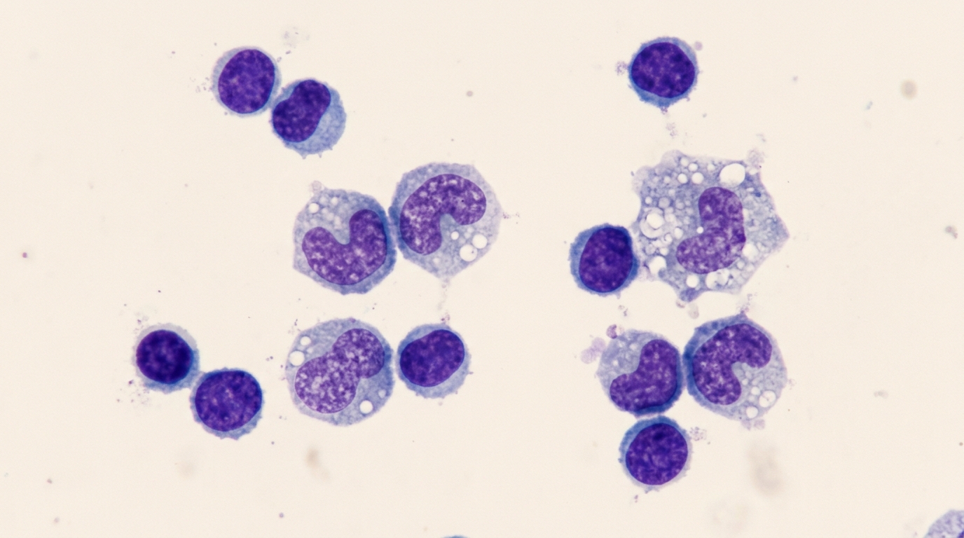
Morphology is where automation falls furthest short. Automated differential counts generally showed weaker agreement, and atypical or malignant cells could not be reliably identified without morphological review. Automated cell differentiation provided by routine analysers is, at best, coarse, distinguishing for example granulocytes, lymphocytes and monocytes, and should be considered for screening only. Automated systems will not reliably recognise the cytomorphological entities that drive differential diagnosis, such as plasma cells, activated lymphocytes, blasts, siderophages, erythrophages, scattered tumour cells or cell clusters. Trained cytologists examining stained cytocentrifuge preparations can detect subtle clues including reactive lymphocytes, plasma cells, activated macrophages, granulocytes, atypical and malignant cells, mitotic figures and bacteria, findings critical in neoplastic and inflammatory disorders.
The review catalogues a range of diagnostic pitfalls that can mislead even careful observers. Artificial blood contamination has been detected in up to 70 percent of all cerebrospinal fluid samples taken, with the incidence appearing even higher in infants and neonates. During lumbar puncture, cells from traversed tissues such as skin, muscle, cartilage, bone marrow and endothelium may appear as artefacts, and endothelial or cartilage cells can be mistaken for pathology. Autolysis begins within hours and first affects granulocytes, producing shadow cells, karyolysis and blurred differential counts. Staining artefacts, including deep-purple reagent granules that mimic bacteria, and cytospin-related mechanical distortion of fragile nuclei add further hazards. The authors also caution that differential counts based on 100 randomly identified cells are point estimates subject to binomial variation: a measured proportion of 30 percent carries an approximate 95 percent confidence interval of 21 to 40 percent, so minor deviations may reflect sampling variability rather than true biological differences.
In inflammatory disease, the review stresses that cytological patterns are not disease-specific and cannot provide aetiological assignment on their own. Acute purulent bacterial meningitis typically produces high cell counts, usually exceeding several thousand cells per microlitre, with neutrophils constituting more than 90 percent of leukocytes in classic adult presentations. Yet infections progress in phases: in the early phase of bacterial meningitis, roughly within 12 hours, bacteria may be directly detectable alongside a lower inflammatory response, and the cell count may appear normal or near normal, sometimes without neutrophilic infiltration, a particularly threatening form known as apurulent meningitis that occurs more frequently in elderly and immunocompromised patients. In severely immunocompromised patients, lower pleocytosis or even normal leukocyte counts may occur, so a normal count does not rule out infection. Viral infections usually show lymphocytic pleocytosis of typically 50 to 500 cells per microlitre by the time of diagnostic puncture, though for several virus types the findings may transiently lack pleocytosis or show a variable neutrophilic skew.
Haemorrhage detection is presented as a uniquely cytological strength. Cranial computed tomography may be unremarkable in a relevant proportion of patients with subarachnoid haemorrhage, especially in subacute scenarios, and targeted cytopathological analysis then provides decisive evidence. After bleeding, macrophages that phagocytose blood components appear in a time-dependent sequence: erythrophages can usually be observed within 4 to 12 hours after the event, gradually evolving into siderophages, which usually occur after 24 to 48 hours and contain granular dark hemosiderin that stains positively with Prussian blue. Later, characteristic yellow-orange rhomboid or star-like haematoidin crystals, the bilirubin crystals, can be detected within siderophages and may float in the fluid for months after macrophage apoptosis, becoming clearly visible under polarised-light microscopy. The precise morphological classification of these phagocytic cells allows conclusions about the temporal stage or acuity of bleeding, though reappearance of erythrophages in sequential samples raises concern for repeated or persistent bleeding and demands correlation with clinical course and imaging.
In neoplastic disease, the authors emphasise that a normal cerebrospinal fluid leukocyte count does not exclude central nervous system neoplasia, and guideline groups advise performing cytology whenever clinical or MRI findings keep pretest probability high. The appearance of malignant cells is highly variable, and no single cytological parameter is pathognomonic for malignancy; the more malignancy criteria found, such as abnormal cell size, enlarged polymorphic nuclei, atypical mitoses and high nucleus-to-cytoplasm ratio, the more reliable the diagnosis. Pre-analytical deterioration can itself mislead, since progressive nuclear fading in autolytic specimens may produce an apparent pseudo-nucleolus mimicking malignancy. When cytology is positive, morphology alone rarely permits definitive tumour classification, prompting adjunct techniques such as immunocytochemistry, flow cytometry or biopsy. Across cancers, the first tap’s sensitivity of roughly 50 to 60 percent rises to about 80 percent with repeat collections, underlining the recommendation to obtain large volumes, process promptly and repeat the puncture if the first specimen is negative but suspicion persists.
The emerging digital layer is treated with measured optimism. Over the past five years, deep-learning systems have moved from early proof-of-concept approaches to small clinical feasibility studies: convolutional models trained on digitised cytospins can perform five-way leukocyte and tumour-cell differentials and even subtype carcinoma cells by putative primary site, with a 2022 pipeline reporting robust cancer detection and subtype classification on more than 50,000 single-cell crops from 90 patients with leptomeningeal metastasis. Evidence is stronger in other body fluids, especially pleural effusions, where artificial intelligence on whole-slide images has reached diagnostic-grade performance for malignancy detection in retrospective cohorts. Regulated AI triage already exists in cytology, with the Hologic Genius platform holding FDA clearance for pap smears, providing a regulatory precedent that may become transferable to body-fluid cytology. Telecytology, delivered through remote-controlled live microscopy or whole-slide imaging, offers a practical way to centralise expertise at peripheral sites, though routine digitisation of paucicellular cerebrospinal fluid cytospins remains technically challenging because autofocus algorithms may fail on sparsely distributed cells. The authors conclude that these studies remain single-centre, often with curated datasets and limited external validation, so generalisability across laboratories, stains and scanners is not yet assured, and all such tools should operate under the oversight of a trained cytopathologist.
Subject of Research: Diagnostic principles, pitfalls and emerging digital tools in cerebrospinal fluid cytology
Article Title: CSF cytology: core principles, diagnostic pitfalls and emerging digital tools including artificial intelligence and telecytology
Article References: Nay, S., Konen, F. F., Bachhuber, F., Dressel, A., Erhart, D., Gerritzen, A., Gross, C. C., Harrer, A., Hinger, A., Lewczuk, P., Lewerenz, J., Khalil, M., Korporal-Kuhnke, M., Leypoldt, F., Möllenkamp, T. M., Otto, M., Petersmann, A., Regeniter, A., Roskos, M., … the German Society for Cerebrospinal Fluid Diagnostics, Clinical Neurochemistry (DGLN e.V.) (2026). CSF cytology: core principles, diagnostic pitfalls and emerging digital tools including artificial intelligence and telecytology. Acta Neuropathologica, 152(1), Article 64. https://doi.org/10.1007/s00401-026-03101-7
Image Credits: AI Generated
DOI: 10.1007/s00401-026-03101-7
Keywords: cerebrospinal fluid, cytology, leukocyte count, automated cell counting, artificial intelligence, telecytology, subarachnoid haemorrhage, neoplastic meningitis, pre-analytical factors, diagnostic pitfalls, cytospin, deep learning
News Source: Ophelia Keating. (November 6, 2026). CSF cytology endures as AI and telecytology reshape spinal fluid diagnostics. Scienmag.