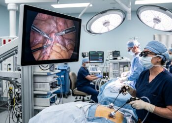
Scientists have built an inferior vena cava filter that lights up under magnetic resonance imaging, and they have shown in a multistage preclinical study that it can be implanted and retrieved in pigs entirely under MRI guidance at 3 Tesla. The work, published in CVIR Endovascular, addresses a long-standing obstacle: conventional filter procedures rely on fluoroscopy, which exposes patients and staff to ionizing radiation and requires iodinated contrast agents. An MR-resonant filter, by contrast, could be placed and removed while the interventionalist watches the device, the vessel anatomy, and even thrombus burden directly on the MRI console.
Inferior vena cava filters are established minimally invasive devices for preventing venous thromboembolism in patients who cannot take anticoagulants or who suffer recurrent embolic events. The filter is threaded through the veins into the large vessel that returns blood from the lower body to the heart, where it traps migrating clots. Retrieval is equally important, because filters left in place too long can become difficult to remove when fibrin deposition or thrombus formation obscures the retrieval hook. Under fluoroscopy, that hook may be hard to see; under MRI, the research team argues, the filter position and hook accessibility can be visualized directly, potentially allowing image-based retrieval planning and removal within the same session.
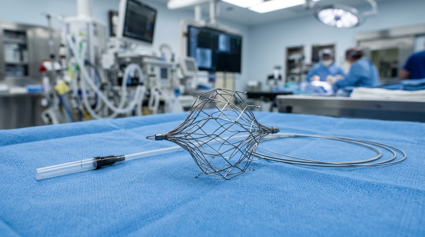
The new device, developed at the Innovation Center Computer Assisted Surgery in Leipzig with manufacturing partners in Germany, is cut from nitinol tubing and expanded to a final diameter of 30 millimeters. Its trick is resonance. Electrically insulated loop-antenna structures with integrated capacitance are tuned to the proton Larmor frequency at 3 Tesla, 128 megahertz, so the filter itself amplifies the local radiofrequency signal and appears bright on the image. The design required electrically separated strut components to prevent short-circuiting between resonant elements, with mechanical integrity maintained by medical-grade adhesive junctions at the proximal and distal filter ends. A companion snare loop, expanded to 20 millimeters and built on the same resonant principle, was designed to capture the filter hook during retrieval.
The evaluation followed a three-stage framework. Stage I characterized device visibility in a saline phantom using static balanced steady-state free precession imaging on a 3 Tesla Biograph mMR system. The filter was oriented parallel to the main magnetic field, matching its expected anatomical orientation in the vena cava, and measurements were taken at flip angles of 10, 20, and 40 degrees and after rotations of the device around its long axis at 22.5, 45, 67.5, and 90 degrees. A single filter was tested at two constrained diameters, 13 and 20 millimeters, representing different implantation states. Quantitative analysis based on ASTM F2119 criteria used a custom Python tool to extract signal profiles across the devices.
The statistics told a clear story. An additive ordinary least-squares model with type II ANOVA found significant effects of flip angle, rotation angle, and filter diameter on normalized signal intensity. Flip angle dominated, with an F value of 275.71 and an effect size of 0.96, followed by rotation angle at F 15.13 and effect size 0.73, while diameter showed only a moderate influence at F 5.22 and effect size 0.19. The strongest signal enhancement occurred at a flip angle of 10 degrees, which the authors attribute to local radiofrequency field amplification by the resonant circuit raising the effective flip angle at the device location. Normalized signal maxima above the predefined detection threshold of 1.3 appeared for most rotation and flip angle combinations; only two configurations fell below it, both involving the 20 millimeter filter at 40 degrees flip angle.
The snare loop behaved differently. In its expected in vivo retrieval orientation, with the loop plane parallel to the transverse imaging plane, no signal enhancement exceeded the threshold, although visibility within the loop area remained preserved. When the loop was turned into the sagittal plane, resonant enhancement did exceed the criterion. The authors note that signal visibility inside the loop interior was nevertheless sufficient for reliable positioning and engagement of the filter hook under MRI guidance, while acknowledging that further optimization of the resonant loop geometry will be required. In vivo, the snare itself showed no resonant enhancement, so positioning at the caudally oriented filter hook was performed using a catheter equipped with passive iron-oxide markers.
Stage II moved the work into a polymer-based vessel phantom built from CT data of the Visible Human dataset, with physiological pulsatile flow simulated by a saline-glycerol mixture and a centrifugal pump. The delivery system, which carries a semi-active MR-resonant saddle coil marker, was advanced over an MR Conditional guidewire and positioned under continuous visualization before the filter was deployed by sheath pullback and a fiberglass pusher rod. Femoral access was established under ultrasound guidance outside the scanner room. As a qualitative test of thrombus interaction, an in vitro thrombus prepared from coagulated porcine blood was flushed through the phantom toward the deployed filter and was successfully captured, with the hypointense thrombotic material visible on the image extending cranially from the device.
Stage III brought the system into five female pigs with a mean body weight of 47.4 kilograms, as part of a larger ethically approved animal study of MRI-guided cardiovascular interventions. The operators had prior catheter-based interventional experience and had practiced the procedure in the phantom, but no prior clinical experience with IVC filter procedures. Initial implantation succeeded on the first attempt in all five animals. In three animals, the deployed filters were retrieved with the snare loop and then re-implanted, achieving retrieval and re-implantation in all three retrieval attempts and bringing the total to eight successful MRI-guided implantations. Retrieval was not attempted in the first two experiments, which were dedicated to sequence and workflow optimization. No structural failure of the resonant filters occurred during delivery or retrieval. Mean implantation time was 8.3 minutes with a standard deviation of 4.8, and mean retrieval time was 13.1 minutes with a standard deviation of 11.1.
Quantitative line-profile analysis confirmed resonant signal enhancement of every deployed filter above the 1.3 threshold in vivo, and the semi-active marker on the delivery system enabled continuous visualization during advancement within the vena cava. The authors emphasize that this work differs from previous MRI-guided IVC filter studies, which were performed predominantly at 1.5 Tesla and mostly demonstrated technical feasibility with conventional off-the-shelf filters. The present study demonstrates a dedicated MR-resonant retrievable filter and snare system within a structured MRI-guided workflow at 3 Tesla, where higher signal-to-noise ratio and stronger resonant amplification may improve device conspicuity, though more careful optimization of sequence parameters and radiofrequency-related device behavior is needed. The workflow also linked device navigation, deployment, and retrieval to dedicated visualization strategies rather than treating feasibility in isolation.
The limitations are spelled out plainly. The study was confined to phantom experiments and acute porcine evaluation; species-specific differences exist between the porcine caudal vena cava and the human inferior vena cava, particularly in the hepatic region. Future studies must assess retrieval performance after long-term implantation, including conditions of fibrin deposition and endothelial incorporation at the filter hook that could complicate removal in patients. Although initial radiofrequency-induced heating experiments were performed, comprehensive safety evaluation under clinically relevant imaging conditions remains necessary. The devices could also be used at 1.5 Tesla, but the integrated capacitance would need retuning to the lower Larmor frequency. The authors position MRI guidance as a specialized investigational approach for now, particularly relevant when radiation or iodinated contrast media are contraindicated, with ultrasound-guided bedside placement remaining an alternative radiation-free option especially in critically ill patients. Still, the structured multistage evaluation, which supported the 3R principles of replacement, reduction, and refinement, may serve as a template for developing other MRI-guided endovascular devices, and it marks a concrete step toward clinically applicable MRI-guided placement and retrieval of inferior vena cava filters.
Subject of Research: Preclinical evaluation of an MR-resonant retrievable inferior vena cava filter and snare loop for MRI-guided implantation and retrieval at 3 Tesla
Article Title: Multistage preclinical evaluation of a retrievable 3 T MR-resonant inferior vena cava filter and snare loop for MRI-guided interventions
Article References: Gholami Bajestani, D., Reich, C. M., Birkigt, J., Mulik, C., Kauffold, J., Sigmarsson, H. L., Wahl, L., Schmid, M., Schauer, S., Jochimsen, T., Sattler, B., Sabri, O., Immel, E., & Melzer, A. (2026). Multistage preclinical evaluation of a retrievable 3 T MR-resonant inferior vena cava filter and snare loop for MRI-guided interventions. CVIR Endovascular, 9(1), Article 118. https://doi.org/10.1186/s42155-026-00770-z
Image Credits: AI Generated
DOI: 10.1186/s42155-026-00770-z
Keywords: MRI-guided intervention, inferior vena cava filter, 3 Tesla MRI, snare loop retrieval, MR-resonant device, preclinical study, porcine model, nitinol filter, interventional radiology, radiation-free imaging, bSSFP imaging, device visibility
News Source: Ophelia Keating. (November 6, 2026). MRI-Guided Blood Clot Filter Shows Promise in Animal Tests. Scienmag.