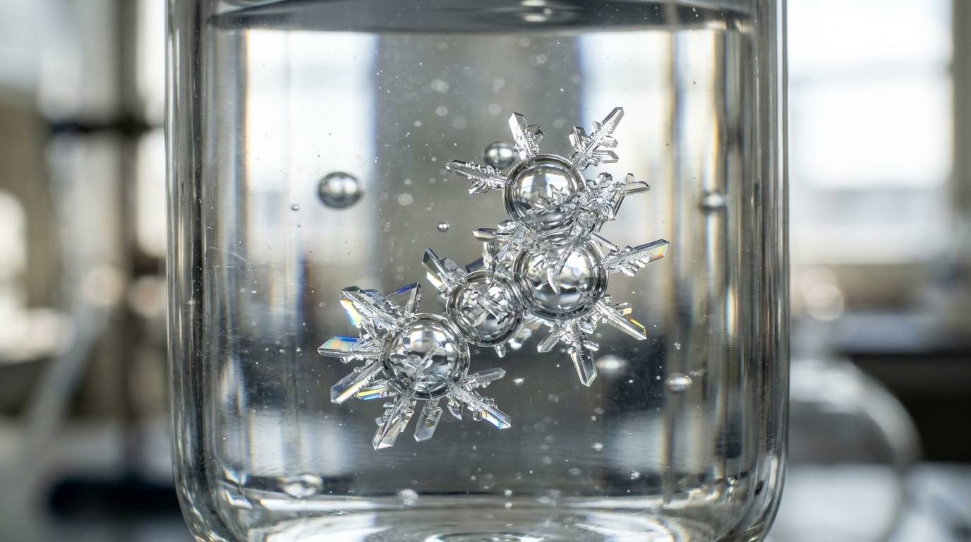
Air pockets too small to see are turning out to be anything but inert passengers in the liquids of the chemistry lab. In a new study published in the Journal of Nanoparticle Research, researchers report that nanobubbles—bubbles under one micron in size, stabilized by surfactant molecules—dramatically accelerate the crystallization of a model mineral from metastable, supersaturated solutions. The work combines spectroscopic, refractometric and light scattering characterization of the bubbles themselves with precipitation experiments, and it concludes that nanobubbles are not passive constituents of chemical solvents but potentially active promoters of the reactions taking place within them.
The study focused on hydroxyapatite, the most sparingly soluble of all pure calcium phosphates, precipitated from solutions whose degree of supersaturation ranged between 14.23 and 15.18 across the pH 7.0 to 7.2 window used in the experiments. These conditions were deliberately chosen to produce nucleation lag times measurable in minutes to hours, so that any accelerating or inhibiting influence of the bubbles would be detectable. Three surfactants of different ionic character were used to stabilize the bubbles: cationic cetyltrimethylammonium bromide, known as CTAB; anionic sodium dodecyl sulfate, or SDS; and nonionic polyoxyethylene sorbitan monolaurate, better known as Tween 20. Each was employed at 80 percent of its critical micelle concentration, a level high enough to give strong spectroscopic signals while keeping the molecules monomeric and avoiding interference from self-associated micellar structures.
The kinetic results were striking. In control solutions containing surfactant but no nanobubbles, the nucleation induction time was 8 ± 1 minutes for both CTAB and SDS, and roughly an hour for Tween 20. When nanobubbles were present, those times collapsed to 1.0 ± 0.2 minutes for CTAB, a mere 5 ± 1 seconds for SDS, and 25 ± 5 minutes for Tween 20. For context, in ultrapure water devoid of any surfactant, irreversible nucleation took around 48 hours. The surfactants themselves were already powerful nucleation promoters, and the degree of promotion tended to increase with surfactant concentration. Because surfactant concentration and chemical identity covaried across the three systems, the authors note that the two effects cannot be fully separated in this design; yet the consistent shortening of the lag time upon bubble introduction, seen for each of the three surfactants, points to the nanobubbles as the source of the additional augmentation.
Controls helped rule out artifacts. Ultrapure water subjected to the same ultrasonication treatment, and ultrasonicated water further purged with nitrogen gas for 60 minutes, both showed nucleation lag times indistinguishable from untreated ultrapure water at approximately 48 hours. Energy-dispersive X-ray analysis detected no significant titanium in any of the nanobubble-assisted precipitates relative to precipitates grown in ultrapure water, addressing the possibility that erosion of the titanium sonication probe had contaminated the chemistry. The authors also caution that the bubble suspensions were characterized within three hours of preparation and their persistence over the full precipitation experiments was not assessed, so the reported effects pertain primarily to the nucleation stage, which occurred within the first hour in all surfactant-containing solutions.
Before any precipitation began, the team characterized the bubbles themselves. Dynamic light scattering placed their hydrodynamic diameters consistently in the 100 to 200 nanometer range regardless of surfactant, a sharp contrast to the 1 to 2 nanometer features detected in the surfactant solutions before sonication. Zeta potential measurements tracked the ionic character of each surfactant: SDS produced the smallest bubbles with a strong negative surface charge, CTAB produced bubbles of similar magnitude but opposite sign, and nonionic Tween 20 yielded the largest bubbles with the lowest absolute zeta potential, averaging around −5 millivolts. The size of the bubbles was inversely related to the absolute value of the zeta potential. The authors stress, however, that light scattering alone cannot establish that the detected objects are gas-filled, and the number density of the bubbles was not determined here.
Nuclear magnetic resonance spectroscopy offered the most intimate structural clues. In proton NMR, the ionic surfactants CTAB and SDS showed consistent downfield shifts of their tail hydrogen protons alongside upfield or insignificant shifts at or near the headgroups, a pattern the researchers interpret as the molecules orienting with their hydrophobic tails toward the gaseous interior of the nanobubbles while the charged headgroups remain immersed in the water phase. Because many shifts were on the order of 0.001 to 0.01 ppm, very near the digital resolution of the instrument, the authors flag this interpretation as partially hypothetical. Tween 20 behaved differently: all hydrogen protons except those of the polyoxyethylene chain shifted upfield, and the POE resonances showed no statistically significant change. The team suggests the nonionic surfactant, immune to charge compensation through hydration and counterion complexation, may penetrate the gaseous milieu inside the nanobubbles even deeper with its tails, though this remains a proposed explanation rather than a settled mechanism.
Infrared spectroscopy and refractometry added complementary evidence. FTIR analysis revealed a consistent decrease in the energy of the hydroxyl stretching mode near 3300 cm−1 when nanobubbles were introduced, contrary to the increase one might expect from reduced hydrogen bonding at a hydrophobic interface. The authors read this as conforming to a model of the interface populated by intense charge separation and a relatively high concentration of free hydroxyls and other transiently immobilized chemical species. They also note the FTIR evidence should be regarded as qualitative and indirect, since water molecules directly at the interface are a minute fraction of the sampled volume. Refractive index, meanwhile, rose slightly in every surfactant solution upon bubble formation—for example, from 1.33340 ± 0.00013 to 1.33344 ± 0.00017 in the CTAB system—despite air having a far lower refractive index than water. The proposed explanation involves charge separation and hydroxide segregation within the Stern layer increasing the total electronic polarizability of the system, though the authors call this interpretation tentative, noting the increases are small and a correlation with bubble concentration was not established.
The precipitates themselves bore the fingerprints of the accelerated nucleation. X-ray diffraction identified poorly crystalline hydroxyapatite as the sole calcium phosphate phase in all seven precipitates, and average crystallite sizes were sub-5 nanometers throughout. Both crystallinity and particle size showed a consistent tendency to decrease when nanobubbles were introduced, with the effect most critical in SDS solutions and least apparent in Tween 20 ones. Electron microscopy quantified the shift: particles grown in CTAB solutions shrank from 148 ± 31 nanometers to 38 ± 11 nanometers on average, SDS platelets fell from tens of microns in length and width to 1.6 ± 0.7 microns, and Tween 20 particles declined from 215 ± 46 to 192 ± 52 nanometers. The authors attribute this not to growth inhibition but to a downstream consequence of promoted nucleation: a shorter lag time means a higher nucleation rate, a greater concentration of nuclei, and therefore smaller, less crystalline crystals sharing the same mass of precipitate.
Surfactants, it turns out, sculpt crystals in a way nanobubbles then undo. Particles from surfactant-containing solutions were invariably larger than the ultrafine 36 ± 7 nanometer particles from ultrapure water, and they adopted rod- and plate-like shapes with aspect ratios rising from 3.2 in water to 3.9, 72 and 11.6 in CTAB, SDS and Tween 20 systems respectively—evidence that surfactants guide growth along preferential crystallographic directions by adsorbing onto specific crystal faces. In the presence of nanobubbles, this anisotropic growth was suppressed, leading the authors to suggest that the bubble interface competes with the amphiphilic molecules and redistributes them away from the growing crystal surfaces. Nanobubbles also raised the equilibrium turbidity of the solutions after about 24 hours, by 12.1 ± 1.2 percent in CTAB, 66.8 ± 1.4 percent in SDS and 9.8 ± 0.6 percent in Tween 20 relative to the corresponding controls. The study concludes that nanobubbles deserve increasing attention for their effects on chemical reactions in the solutions that harbor them, with consequences for nucleation kinetics, crystal growth and the ultimate properties of synthesized materials.
Subject of Research: Surfactant-stabilized nanobubbles and their effects on hydroxyapatite crystallization from metastable supersaturated solutions
Article Title: Physicochemical characterization of surfactant-stabilized nanobubbles and their effect on crystallization from metastable supersaturated solutions
Article References: Uskoković, V. (2026). Physicochemical characterization of surfactant-stabilized nanobubbles and their effect on crystallization from metastable supersaturated solutions. Journal of Nanoparticle Research, 28(11), Article 286. https://doi.org/10.1007/s11051-026-06793-w
Image Credits: AI Generated
DOI: 10.1007/s11051-026-06793-w
Keywords: nanobubbles, surfactants, crystallization, hydroxyapatite, nucleation, supersaturation, NMR spectroscopy, FTIR, refractive index, zeta potential, X-ray diffraction, electron microscopy
News Source: Denise Maddox. (November 5, 2026). Nanobubbles emerge as active promoters of crystal formation in supersaturated solutions. Scienmag.