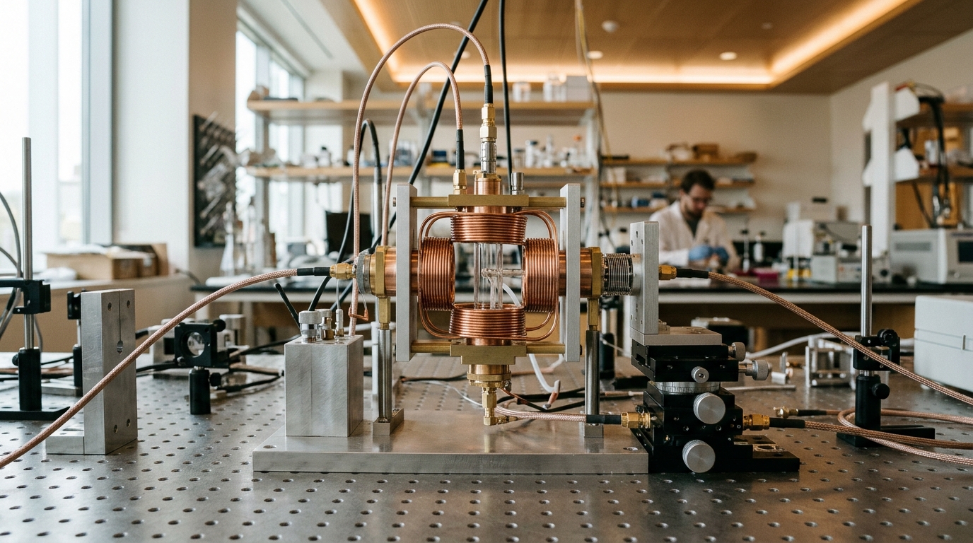
In a study published in Discover Imaging, researchers at the University of Colorado Denver induced acute lung injury in mice with an intraperitoneal dose of lipopolysaccharide (LPS), then used specialized cyclic hydroxylamine probes to trap superoxide in blood, lung cells, and lung mitochondria.
Wild-type C57BL/6J mice and a transgenic strain overexpressing extracellular superoxide dismutase (EC-SOD) specifically in the lung were given a single 10 mg/kg intraperitoneal injection of LPS from E. coli. Twenty-four hours later, the animals received one of three spin probes: CMH delivered intraperitoneally and subcutaneously for blood measurements, or CPH and DCP-AM-H delivered intratracheally for lung cellular and mitochondrial superoxide detection, respectively. Blood was drawn one hour after CMH administration, while lungs were harvested five minutes after intratracheal probe delivery.
EPR spectroscopy is considered the gold standard for detecting, identifying, and quantifying free radicals, but endogenous radical species exist at extremely low steady-state concentrations—superoxide inside cells, for example, sits in the nanomolar range, well below direct detection limits. Spin traps solve this problem by reacting with short-lived radicals to produce more stable, long-lived nitroxide radicals whose distinctive spectra can be measured. The nitroxide signal is proportional to the amount of superoxide trapped during the experiment, minus a decrement caused by bio-reduction of the nitroxide itself, and is used to determine changes in superoxide production rather than absolute concentrations.
To measure blood superoxide, the team used X-band EPR at approximately 9.5 GHz on 50-microliter blood samples in capillary tubes at room temperature. The concentration of the nitroxide CM·, generated when CMH reacts with superoxide, was determined using the Bruker Spin Fit and spin count module. The same LPS exposure also produced clear signs of systemic inflammation: neutrophils and monocytes increased while platelets decreased, consistent with sepsis, alongside a modest but significant decrease in lymphocytes and increases in eosinophils and basophils.
Lung injury itself was confirmed through bronchoalveolar lavage fluid (BALF) analysis. Total protein in the BALF supernatant, measured with a BCA protein assay, rose after LPS treatment, reflecting disruption of the epithelial and endothelial barrier—the hallmark protein leak of ALI. Total alveolar cell counts also increased.
The central technical advance came in the lung imaging experiments. Because X-band microwaves are strongly absorbed by water-laden samples, limiting studies to 8–10 mg of excised tissue or 50 microliters of fluid, the researchers turned to L-band EPR at 1 GHz, where energy absorption is decreased and tissue penetration is greater. Using a rapid-scan EPR technique—in which the magnetic field is scanned through resonance at kHz frequencies, allowing extensive signal averaging and substantial signal-to-noise improvements—the team imaged intact excised lungs placed side-by-side in an 8 mm outer diameter tube. The spectral-spatial images displayed the characteristic 3-line nitroxide EPR spectrum as a function of position along the magnetic field gradient, with no other EPR signals observed.
With the CPH probe, the researchers detected a significant increase in cellular superoxide in lungs from LPS-treated wild-type mice compared with PBS controls. Using DCP-AM-H, a probe that preferentially loads into mitochondria, they similarly detected increased mitochondrial superoxide. Spin counts of CP· and DCP· were obtained from non-gradient spectra acquired before recording each spectral-spatial image. The image resolution was not high enough to distinguish signals from the two closely spaced lungs, but the observation of EPR signals strong enough to image in excised lungs—and the expectation of similar signal strengths in vivo—suggests the feasibility of future imaging in living animals.
The most striking result emerged from the transgenic mice. These animals carry a sod3 transgene driven by the surfactant protein C promoter in type II alveolar epithelial cells, expressing 2.5 to 4 times more lung EC-SOD and previously shown to be protected against lung injury. When these mice received LPS, their blood superoxide still increased, yet lung cellular and mitochondrial superoxide did not rise.
This dissociation carries significant implications for how oxidative stress has been studied in ARDS. Inflammation drives reactive oxygen species production and oxidative stress is a hallmark of acute respiratory distress syndrome, yet antioxidant treatment trials have not proven effective at reducing mortality. A major contributing factor, the authors argue, is that lung redox status was never directly measured in patients; instead, oxidative stress measures have relied on end-point product accumulation in plasma as surrogates for lung ROS. Clinical antioxidant trials used blood markers such as protein carbonyls and MDA plus 4-HNE as proxies for lung redox status, and these trials did not improve outcomes in the populations studied.
The authors are careful to note the limitations of the method. Although hydroxylamine probes are oxidized by superoxide to the corresponding nitroxides, the measured nitroxide concentrations may not exactly mirror superoxide concentrations in situ, because nitroxides are susceptible to bio-reduction—for example by ascorbate—back to the hydroxylamine, which can then be oxidized again. These redox cycling processes complicate interpretation of measured nitroxide as total superoxide produced during the experiment, so the study focuses on changes in superoxide production rather than absolute quantification. Data were analyzed with unpaired t-tests, with significance set at p < 0.05, and sample sizes ranged from 4 to 13 animals per comparison depending on the measurement.
Compared with earlier approaches, the study advances the field in specific ways. HPLC detection of superoxide-specific oxidation products of dihydroethidium has been successful across various organs after LPS treatment but cannot be used for imaging. A previous study demonstrated the feasibility of in vivo CPH injection after LPS exposure with subsequent X-band EPR measurements at liquid nitrogen temperature. The current work instead measures superoxide production in intact tissues at room temperature through low-frequency L-band EPR, building on the team’s own prior protocols for probe delivery and X-band spectroscopy in live mice and for rapid-scan EPR at lower frequencies.
The long-term goal is in vivo EPR imaging of lungs to evaluate lung redox status quantitatively in ARDS, enabling disease risk stratification and guiding clinical research. Recent work on ARDS has highlighted the importance of identifying hyper-inflammatory and hypo-inflammatory patient subphenotypes associated with different therapy responses, and mortality remains high with increasingly clear long-term effects on survivors. Future studies will develop the capacity to monitor lung redox status in live animals directly, moving the measurement from the excised organ to the intact, breathing lung.
Subject of Research: Direct EPR detection and imaging of cellular and mitochondrial superoxide in a mouse model of acute lung injury
Article Title: Electron paramagnetic resonance detection of superoxide in a murine model of acute lung injury
Article References: Elajaili, H. B., Dee, N., Hovey, T., Canny, A., Amassah, G., Posey, J., Rinard, G. A., Kao, J. P. Y., Eaton, S. S., Eaton, G. R., & Nozik, E. S. (2025). Electron paramagnetic resonance detection of superoxide in a murine model of acute lung injury. Discover Imaging, 2(1), Article 11. https://doi.org/10.1007/s44352-025-00014-1
Image Credits: AI Generated
DOI: 10.1007/s44352-025-00014-1
Keywords: electron paramagnetic resonance, superoxide, acute lung injury, LPS, EPR imaging, spin probes, EC-SOD, mitochondria, reactive oxygen species, ARDS, lung redox status, murine model
News Source: Denise Maddox. (November 5, 2026). Superoxide in injured mouse lungs imaged directly by low-frequency EPR. Scienmag.