


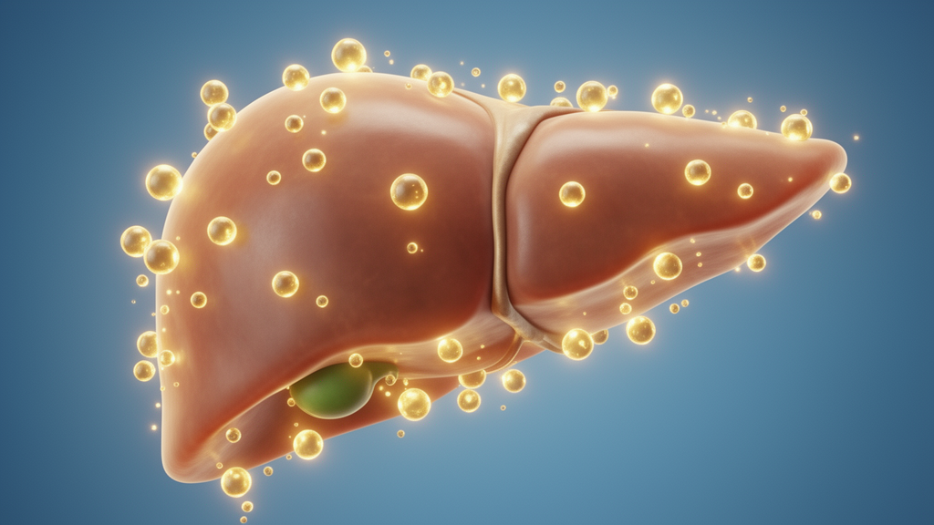
Tauroursodeoxycholic acid, or TUDCA, is a hydrophilic secondary bile acid that has attracted growing attention for its liver-protective properties, and a new narrative review published in Frontiers in Pharmacology brings together what is currently known about how it works and where it might eventually fit into clinical practice. The review, authored by researchers at the First Affiliated Hospital of Xi’an Medical University, synthesizes molecular targets and signaling pathways across metabolic dysfunction-associated steatotic liver disease, cholestatic liver injury, liver fibrosis, hepatocellular carcinoma, hepatic ischemia-reperfusion injury, and gallbladder stones. Its central message is a careful one: preclinical evidence is broad and mechanistically rich, but high-quality clinical data supporting routine use remain limited, and the interplay between TUDCA and the gut microbiota is still poorly understood.
TUDCA is the taurine-conjugated form of ursodeoxycholic acid, a bile acid naturally found in the bile of hibernating bears and long used in traditional Chinese medicine. Its biosynthesis begins with cholesterol, which hepatocytes convert through the classical CYP7A1-mediated pathway, or a CYP27A1-dependent bypass, into chenodeoxycholic acid. Once secreted into the colon with bile, gut bacteria perform a C7-hydroxyl epimerization: bacterial 7α-hydroxysteroid dehydrogenase oxidizes CDCA to 7-ketolithocholic acid, and 7β-hydroxysteroid dehydrogenase reduces that intermediate to UDCA. Reabsorbed via enterohepatic circulation, UDCA is then taurine-conjugated in hepatocytes to yield TUDCA. UDCA itself is FDA-approved for primary biliary cholangitis, and TUDCA’s enhanced water solubility is thought to make it more readily absorbed and utilized by the intestine and liver.
The review’s mechanistic core centers on endoplasmic reticulum stress and the unfolded protein response. The ER handles protein folding, post-translational modification, and lipid biosynthesis, and when stressors such as nutrient starvation, oxidative stress, or viral infection overwhelm its folding capacity, misfolded proteins accumulate, a state known as ER stress. Three transmembrane sensors, PERK, IRE1α, and ATF6, normally held inactive by the chaperone GRP78, are activated when GRP78 dissociates. In a chronic intermittent hypoxia-induced liver injury model, TUDCA significantly downregulated GRP78 and reduced PERK, IRE1α, and ATF6, while reversing overexpression of apoptosis-promoting proteins including CHOP, activated caspase-12, phosphorylated eIF2α, and phosphorylated JNK, thereby blocking the transmission of stress signals toward cell death.
Beyond ER stress, TUDCA suppresses inflammation, oxidative damage, and apoptosis through interlocking pathways. ER stress activates inflammatory signaling via JNK-AP-1, NF-κB, and IκB kinase-dependent routes, and can trigger the hepatic NLRP3 inflammasome, causing hepatocyte death; TUDCA prevents this by inhibiting ER-dependent inflammasome activation. In a tunicamycin-induced rat model of hepatotoxicity, TUDCA downregulated cytoplasmic phospholipase A2 and cyclooxygenase-2, inhibited prostaglandin E2 production, and restored serum ALT, AST, and total bilirubin levels. In a mouse NASH model, it dampened an lncRNA-driven NF-κB and JNK cascade and reduced TNF-α, IL-6, and IL-1β. In fatty liver ischemia-reperfusion injury, it reduced mitochondrial calcium overload in hepatic macrophages, lowering ROS and suppressing NLRP3 activation, and in obese mice it promoted NRF2 nuclear translocation by inhibiting KEAP1-NRF2 interaction, boosting antioxidant genes such as Nqo1, Ho-1, and Gpx4.
Anti-apoptotic effects have been documented in primary rat hepatocytes exposed to glycochenodeoxycholic acid, where TUDCA reduced caspase-9 and caspase-3 activation through p38 MAPK, ERK1/2, and PI3K signaling, an effect largely independent of NF-κB. Interestingly, neuronal models add complementary detail: in amyloid-β-treated PC12 cells TUDCA suppressed the E2F-1/p53/Bax pathway, and in primary cortical neurons protection involved PI3K/Akt signaling and reduced Bax translocation. The review notes, however, that the contribution of some of these extrahepatic mechanisms to hepatoprotection remains to be clarified.
In metabolic dysfunction-associated steatotic liver disease, now the world’s most common chronic liver disease affecting nearly one-quarter of adults globally, TUDCA showed antisteatotic and lipid-lowering effects in multiple animal models. It reduced long-chain fatty acid uptake by inhibiting FXR and FATP5 expression, and in an HFD-induced model it partially restored gut microbial composition, increasing potentially anti-inflammatory taxa such as Faecalibacterium and Akkermansia while reducing Mucispirillum and Ruminococcus gnavus, alongside improved intestinal barrier function and reduced hepatic steatosis. Preliminary clinical findings align with these observations: in a 24-week study of 186 patients with NAFLD under the previous nomenclature, TUDCA combined with polyene phosphatidylcholine produced greater reductions in liver enzymes and controlled attenuation parameter than glutathione plus phosphatidylcholine, and an earlier randomized study of 180 patients reported improvements in biochemical indices, lipid profiles, and ultrasonographic findings. The authors caution, however, that combination therapy and surrogate endpoints limit conclusions about independent and long-term efficacy.
The strongest clinical dataset concerns cholestatic disease, specifically primary biliary cholangitis. In a multicenter, randomized, double-blind trial of 199 patients, both TUDCA and UDCA at 250 mg three times daily for 24 weeks achieved an alkaline phosphatase reduction greater than 25 percent in 75.97 percent and 80.88 percent of patients respectively, with comparable biochemical responses and safety. The review interprets this as evidence of comparable short-term biochemical efficacy rather than superiority of either agent. Experimental cholestasis models add mechanistic depth: in cholic acid-induced cholestasis, TUDCA increased nuclear FXR and NRF2 levels, enhanced BSEP and MRP2 expression, and reduced NTCP and CYP7A1, while retaining protection in Fxr-deficient mice through NRF2-dependent signaling, indicating it is not simply a uniform FXR agonist or antagonist. In both CA and ANIT models it reduced GRP78 and CHOP and inhibited the CHOP-DR5-caspase-8 apoptotic pathway.
In fibrosis, ischemia-reperfusion injury, and cancer models, the evidence remains preclinical. TUDCA reduced collagen deposition and α-SMA expression in CCl4-treated rats, promoted hepatocyte proliferation in a GATA3-dependent manner, and reduced fibrosis and CHOP expression in a common bile duct ligation model of secondary biliary fibrosis. In hemorrhagic shock and warm hepatic IRI models, it lowered ALT and AST, modulated PERK signaling, suppressed Kupffer cell cytokine release via the IRE1α/TRAF2/NF-κB pathway, and restored SIRT1-FXR signaling. In Mst1/2-deficient and DEN-induced mouse models of hepatocarcinogenesis, TUDCA reduced tumor formation by promoting YAP nuclear export and degradation, and in HCC-bearing mice, Akkermansia muciniphila-enhanced anti-PD-1 responses were accompanied by increased circulating TUDCA, though a proposed FXR-SHP-PD-L1 link was not established. In a murine cholesterol gallstone model, TUDCA reduced stone formation by downregulating ABCG5/ABCG8 and increasing Cyp27 expression.
The review is candid about its limitations. Experimental doses vary enormously, from 2.5 to 1,000 mg/kg/day in high-fat diet models, with in vitro concentrations spanning tens of micromolar to millimolar ranges, making cross-study comparison and clinical extrapolation difficult. Human pharmacokinetic data come mainly from PBC or biliary disposition studies, and microbial deconjugation of TUDCA, as reported for Campylobacter jejuni in an aging-related model, may alter its activity in ways not yet characterized. Clinical evidence outside PBC relies on small trials with short-term biochemical or imaging surrogate endpoints, and some fatty liver studies used combination regimens that obscure TUDCA’s independent contribution. The authors conclude that future studies must move beyond short-term biochemical responses to determine whether experimental benefits translate into sustained, clinically meaningful outcomes, and how TUDCA compares with UDCA and established therapies in specific liver diseases.
Subject of Research: Hepatoprotective mechanisms of tauroursodeoxycholic acid and its therapeutic potential across liver diseases
Article Title: Hepatoprotective mechanism of tauroursodeoxycholic acid and its therapeutic potential in liver diseases
Article References: Chao, R., Zhang, Y., Hao, Y., Zhang, H., Zhang, M., & Sun, C. (2026). Hepatoprotective mechanism of tauroursodeoxycholic acid and its therapeutic potential in liver diseases. Frontiers in Pharmacology, 17, Article 1949645. https://doi.org/10.3389/fphar.2026.1949645
Image Credits: AI Generated
DOI: 10.3389/fphar.2026.1949645
Keywords: TUDCA, bile acids, liver disease, endoplasmic reticulum stress, unfolded protein response, MASLD, primary biliary cholangitis, liver fibrosis, hepatocellular carcinoma, gut-liver axis, oxidative stress, gut microbiota
News Source: Ophelia Keating. (October 29, 2026). Bile Acid TUDCA Shows Liver Protection Across Diseases, But Human Evidence Remains Thin. Scienmag.



