


Cerebrospinal fluid, the clear plasma ultrafiltrate that bathes the brain and spinal cord, has long been one of clinical neurology’s most informative diagnostic windows, yet its value depends entirely on how the sample is taken, handled, and read. A new narrative review published in Discover Neuroscience synthesizes the physiology of cerebrospinal fluid, the technical conduct of lumbar puncture, and a structured, phase-based framework for interpreting cerebrospinal fluid biomarkers across emergency, basic, and extensive diagnostic tiers. Written by Edy Alyson Costa Ribeiro, Leila Antonangelo, and Hélio Rodrigues Gomes, the review draws on PubMed/MEDLINE and SciELO literature, supplemented by Google Scholar, with preference given to publications from the last ten years and to guidelines from international societies including the European Federation of Neurological Societies, the Infectious Diseases Society of America, and the European Federation of Clinical Chemistry and Laboratory Medicine.
The physiological groundwork matters because every downstream measurement depends on it. Cerebrospinal fluid is produced primarily by the ependymal cells of the choroid plexuses at roughly 20 to 25 milliliters per hour, totaling about 500 milliliters per day, while the average circulating volume in an adult stands at only 125 to 150 milliliters. That mismatch means the entire complement is renewed three to four times daily. The fluid flows through the lateral, third, and fourth ventricles and caudally toward the spinal cord before draining via cervical lymph nodes or entering the glymphatic system, ultimately reaching the venous system. Functionally, it absorbs external impacts, supplies nutrients, and removes toxic metabolites from brain tissue.
Barrier biology explains which substances appear in the fluid and in what proportions. The blood-cerebrospinal fluid barrier is composed of ependymal cells surrounding clusters of fenestrated capillaries, with filtration mediated by sodium-potassium ATPase pumps and aquaporins that modulate osmotic gradients. Large molecules such as cells and certain proteins are excluded, while ions and small solutes pass easily. Proteins, unlike glucose, amino acids, lactate, and vitamin C, move mainly through passive molecular diffusion governed by non-linear concentration gradients that depend on molecular size and fluid flow rate. The blood-brain barrier itself consists of endothelial cells joined by tight junctions, embedded within a neurovascular unit that also includes astrocytes, neurons, microglia, and an acellular basement membrane. Permeability is dynamically regulated by calcium signaling, phosphorylation, and the actin cytoskeleton; altered phosphorylation of occludin and ZO-1 can trigger junction disassembly and increased permeability. Breakdown of this barrier is implicated in Alzheimer’s disease, Parkinson’s disease, ALS, stroke, traumatic brain injury, multiple sclerosis, HIV, and meningitis.
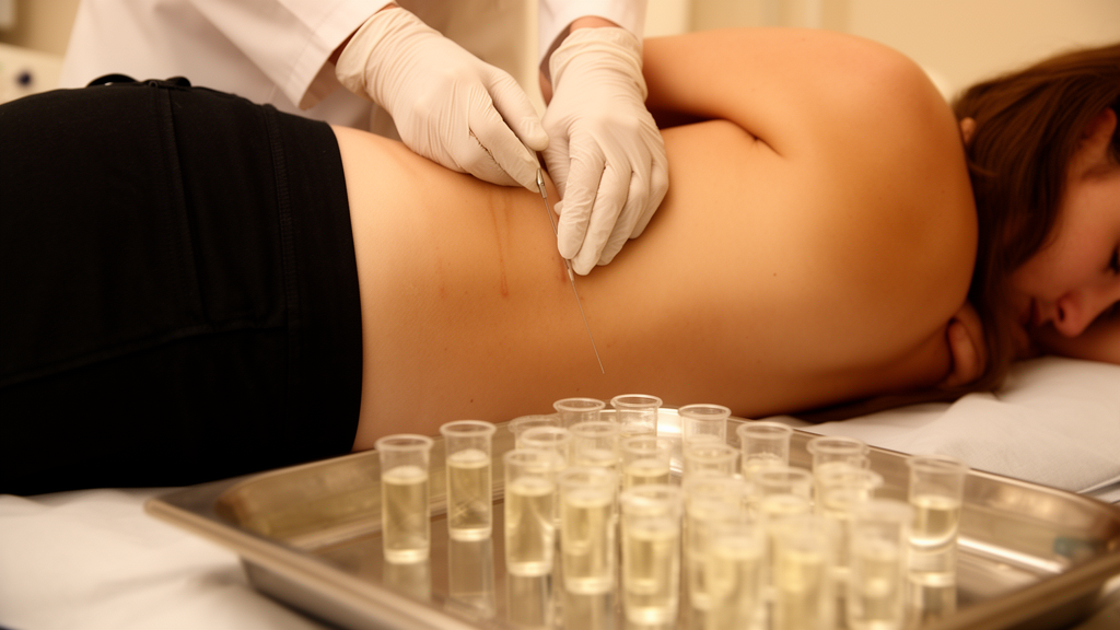
Lumbar puncture remains the gateway to all of this analysis, and the review is unusually specific about technique. The procedure withdraws fluid from the subarachnoid space for diagnostic or therapeutic purposes and for measuring intracranial pressure, with indications spanning meningitis, encephalitis, myelitis, Guillain-Barré syndrome, neuromyelitis optica, subarachnoid hemorrhage, headache differential diagnosis, dementia syndromes, and autoimmune central nervous system disorders. Required materials include sterile gloves, 2% chlorhexidine for skin antisepsis, 5 to 10 milliliters of 1% lidocaine without vasoconstrictor, and a standard 22-27G Quincke-type spinal needle or, alternatively, a pencil-point atraumatic needle to reduce bleeding and post-dural puncture headache. Polypropylene tubes are mandatory because glass or polystyrene tubes adsorb hydrophobic peptides, artificially reducing amyloid-beta 42 concentrations by up to 30%.
Positioning and anatomical landmarks determine both safety and measurement quality. The seated position, leaning forward over a pillow, is acceptable but unsuitable for manometry; the lateral decubitus position with maximal flexion of neck, knees, and hips is preferred for accurate opening pressure. Tuffier’s line identifies the L4 spinous process, and puncture is typically performed at the L3-L4 or L4-L5 interspace. The needle is advanced through skin, subcutaneous tissue, the supraspinous and interspinous ligaments, ligamentum flavum, epidural space, dura mater, and arachnoid membrane, with a distinct loss of resistance marking dural penetration. When opening pressure is indicated, it must be recorded before collection; the adult physiological reference range is 6 to 20 centimeters of water, and clinicians must account for confounders such as obesity, increased abdominal pressure, coughing, the Valsalva maneuver, or excessive knee flexion. Abnormal readings point toward distinct states, from spontaneous cerebrospinal fluid leaks causing intracranial hypotension to idiopathic intracranial hypertension, space-occupying lesions, hydrocephalus, meningeal infections, and cerebral venous sinus thrombosis.
The pre-analytical phase, the review emphasizes, is the step with the highest probability of error and the most critical source of diagnostic error and inter-laboratory variability. Fluid is collected into three or four tubes: the first is reserved for chemical and serological testing, while subsequent tubes go to microbiological and cytological analyses to ensure reliable cell counts and minimize bacterial contamination from skin flora. Approximately 0.5 to 1 milliliter suffices for Gram stain and routine culture, while larger volumes of 5 to 10 milliliters increase sensitivity for mycobacteria and fungi. Samples for cell counting and cytology must be processed within one to two hours at room temperature, because delayed processing leads to rapid leukocyte lysis, with up to 50% cell loss within two hours, and in vitro metabolic activity that artificially depresses glucose and elevates lactate. For biochemical and biomarker work, fluid should be centrifuged at 2,000 times gravity for ten minutes at 4 degrees Celsius, preferably within four hours, then aliquoted into low protein-binding polypropylene tubes and stored at minus 80 degrees Celsius, avoiding repeated freeze-thaw cycles. Cytological and microbiological specimens should never be refrigerated, since some clinically important pathogens lose viability at low temperatures.
The emergency tier of interpretation targets acute inflammation and hemorrhage through macroscopic appearance, global cell counts, glucose, and lactate. Physiological fluid is clear and colorless; xanthochromia, a yellowish discoloration from erythrocyte degradation and bilirubin production, appears after six hours in approximately 20% of subarachnoid hemorrhages and in 90% of patients after twelve hours, persisting up to two weeks. A diagnostic threshold of more than 3,667 erythrocytes per microliter, established for the first day post-hemorrhage, yields 97% sensitivity and 94% specificity for subarachnoid hemorrhage, and xanthochromia independently predicts the condition with an odds ratio of 15.5. Conversely, a global cell count below 2,000 cells per microliter without xanthochromia effectively excludes the diagnosis. Pleocytosis patterns vary by pathogen: Streptococcus pneumoniae averages 1,842 cells per microliter, Neisseria meningitidis 5,238, and Haemophilus influenzae type b 1,470. Lactate, reflecting local anaerobic glycolysis and independent of blood concentrations, is a superior marker for differentiating bacterial meningitis, above 3.5 millimoles per liter, from viral meningitis, with the range of 1.2 to 2.1 millimoles per liter most widely accepted for adults and children. Hypoglycorrhachia, defined as glucose below 2.5 to 2.8 millimoles per liter or a cerebrospinal fluid to serum ratio below 0.50 to 0.60, occurs in bacterial meningitis through altered glucose transporters and leukocyte-mediated glycolysis.
The basic tier evaluates barrier integrity and intrathecal inflammation. Approximately 80% of proteins in physiological fluid derive from the systemic circulation, with albumin serving as the primary reference protein because its presence is exclusively systemic. The cerebrospinal fluid to serum albumin quotient remains the most reliable parameter for assessing barrier function. Quotient diagrams, or Reibergrams, distinguish whether elevated immunoglobulins reflect reduced fluid flow or true intrathecal synthesis. IgG oligoclonal bands, evaluated by isoelectric focusing with immunofixation, constitute the gold standard for detecting intrathecal antibody production across five patterns; a Type 2 pattern, bands exclusive to the fluid, is classic in multiple sclerosis and neuroborreliosis, and detection yields 88 to 98% sensitivity for multiple sclerosis diagnosis. In MOG antibody-associated disease, oligoclonal bands appear in only 6 to 22% of cases, against roughly 70% positivity in neuroborreliosis. Adenosine deaminase, with a cutoff above 10 units per liter, aids tuberculous meningitis diagnosis, though pooled accuracy data show sensitivity of roughly 88 to 93% and specificity of 89 to 92% with considerable heterogeneity, and elevation also occurs in lymphoma, bacterial meningitis, and neurobrucellosis.
The extensive tier extends into neurodegeneration, and the review highlights Alzheimer’s disease biomarkers as its representative example. In a longitudinal cohort study of Han Chinese individuals, amyloid-beta 42 concentrations began to diverge from controls 18 years before clinical disease onset, followed sequentially by changes in the amyloid-beta 42 to amyloid-beta 40 ratio at 14 years, phosphorylated tau 181 at 11 years, and total tau at 10 years before diagnosis. As cognitive impairment progresses, these cerebrospinal fluid alterations initially accelerate before decelerating in later disease stages. Beyond these assays, flow cytometry immunophenotyping can characterize immune and hematopoietic cells by antigen co-expression, detect neoplastic infiltration, and resolve up to 95% of samples deemed inconclusive by traditional cytology, proving more sensitive than conventional cytology for blast infiltration in acute leukemia. The authors conclude that rigorous lumbar puncture technique, systematic pre-analytical care, and structured phase-based interpretation together position cerebrospinal fluid analysis as an increasingly critical tool for diagnosing infectious, inflammatory, and neurodegenerative central nervous system disease.
Subject of Research: Cerebrospinal fluid analysis in clinical neurology, from physiology and lumbar puncture technique to biomarker interpretation
Article Title: Cerebrospinal fluid analysis from physiological principles and lumbar puncture to analytical interpretation in clinical neurology
Article References: Ribeiro, E. A. C., Antonangelo, L., & Gomes, H. R. (2026). Cerebrospinal fluid analysis from physiological principles and lumbar puncture to analytical interpretation in clinical neurology. Discover Neuroscience, 21(1), Article 101. https://doi.org/10.1186/s13064-026-00329-7
Image Credits: AI Generated
DOI: 10.1186/s13064-026-00329-7
Keywords: cerebrospinal fluid, lumbar puncture, blood-brain barrier, oligoclonal bands, Alzheimer's disease biomarkers, meningitis, xanthochromia, subarachnoid hemorrhage, opening pressure, pre-analytical phase, lactate, glucose
News Source: Cassandra Pierce. (October 29, 2026). Cerebrospinal Fluid Analysis: From Lumbar Puncture Technique to Clinical Biomarker Interpretation. Scienmag.



