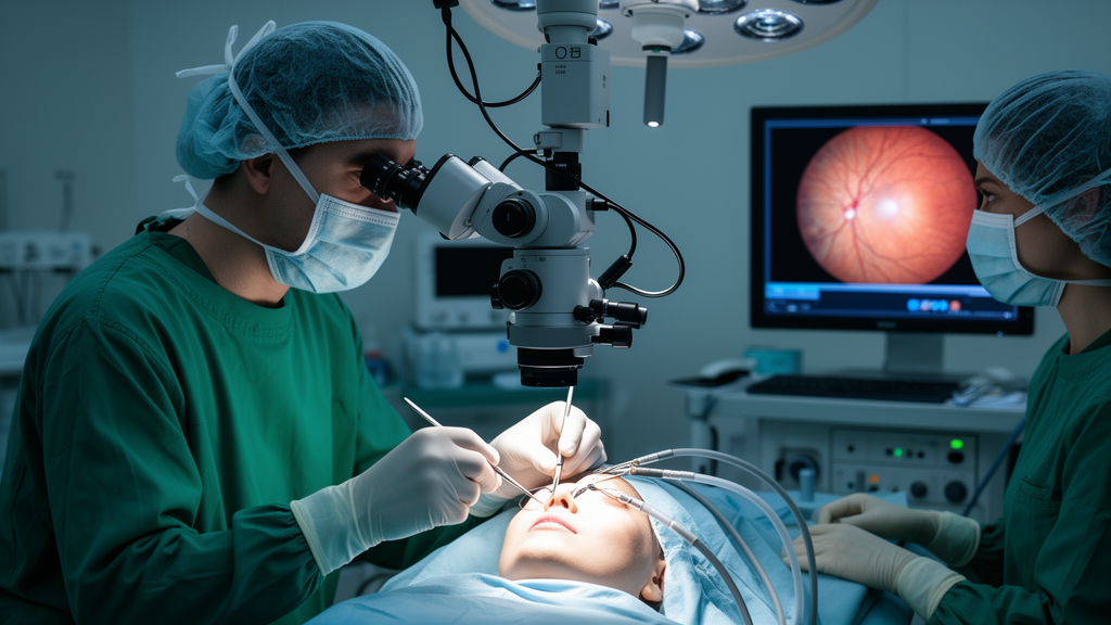
A 52-year-old woman with severe nearsightedness regained limited but stable vision after surgeons in Xiamen, China, repaired a macular hole retinal detachment using a technique that avoids cutting away one of the eye’s most delicate membranes. The retina reattached, the hole closed, and the result held for a full year.
The case, reported in Frontiers in Ophthalmology by Zhi Zhong, Shuxin Cai and Haibo Li of Xiamen Eye Center and Eye Institute of Xiamen University, is a single-patient account and carries all the limits that implies. The authors themselves stress that the relative contributions of the epiretinal proliferation flap and the preserved internal limiting membrane cannot be separated in one eye, and that larger prospective studies are needed before the approach can be recommended more broadly. Still, the report documents a technically feasible way to reconstruct a large myopic macular hole without harvesting donor tissue from elsewhere in the eye or the body.
In highly myopic eyes, the globe elongates, the retina thins, and the posterior pole bulges outward into areas called posterior scleral staphylomas. These changes create a biomechanical environment that works against both macular hole closure and retinal reattachment. The standard operation is pars plana vitrectomy with peeling of the internal limiting membrane, a translucent basement membrane on the inner retinal surface whose removal releases tangential traction on the hole. But even with that traction relieved, anatomical and visual outcomes remain less favorable in highly myopic eyes than in idiopathic macular holes, particularly when myopic degeneration is advanced.
To improve results in difficult holes, surgeons developed the inverted internal limiting membrane flap, in which a piece of the membrane is peeled around the hole’s rim and folded over the fovea to act as a scaffold for glial closure. That technique has substantially improved closure and reattachment rates and is now regarded as the preferred approach for large or complex myopic macular holes. When the membrane is too thin or absent, surgeons have turned to other tissue-based strategies: free internal limiting membrane flaps, lens capsule transplantation, human amniotic membrane grafting, and autologous retinal transplantation. Each of these demands additional donor tissue, extra surgical manipulation, or exogenous material, and each adds complexity to an already delicate operation.
Epiretinal proliferation offers a different kind of scaffold, because it is already sitting on the pathological site. Unlike a conventional epiretinal membrane, epiretinal proliferation is closely adherent to the retinal surface and can be mobilized as a pedicled flap while keeping its retinal attachment, with no tissue harvesting required. Müller cell-derived processes and transdifferentiated Müller cells contribute to such epiretinal tissue and may serve as a substrate during retinal remodeling. The authors caution, however, that preserving this tissue is not universally beneficial. Reactive Müller cell gliosis after retinal injury can be neuroprotective, stabilizing retinal architecture and promoting wound repair, but excessive activation may drive glial scar formation, abnormal remodeling, and persistent structural change. The potential advantage depends on controlled remodeling rather than unchecked proliferation.
The patient in this report illustrates how demanding the anatomy can be. She had progressive blurred vision in her right eye for five months and had undergone cataract surgery with intraocular lens implantation in both eyes ten years earlier. She had no history of ocular trauma, glaucoma, diabetic retinopathy, or previous vitreoretinal surgery, and her systemic medical history was unremarkable. Because she had not had regular ophthalmic follow-up, her vision before symptoms began was never documented. Her left eye saw 20/160. Axial length in the right eye measured 33.89 millimeters, confirming high myopia.
Imaging revealed the full extent of the problem. Fundus examination showed a large full-thickness macular hole with a localized retinal detachment involving the posterior pole, together with distinct superior and inferior posterior scleral staphylomas that gave the back of the eye an irregular configuration. Spectral-domain optical coherence tomography measured the hole’s minimum diameter at 687 micrometers and showed extensive epiretinal proliferation along its margin. The localized detachment corresponded to the superior staphyloma. The left eye, separately, had a lamellar macular hole with an epiretinal membrane. After informed consent, the team proceeded with a 27-gauge pars plana vitrectomy using the inverted epiretinal proliferation flap technique with preservation of the internal limiting membrane.
The operation itself was deliberately conservative. After core vitrectomy and complete posterior hyaloid separation, residual cortical vitreous was carefully removed. Indocyanine green dye was applied during surgery to confirm that the internal limiting membrane remained intact and to help the surgeons distinguish the epiretinal proliferation from the membrane beneath it. The proliferation surrounding the hole was then meticulously dissected into a continuous pedicled flap that kept its retinal attachment, and the flap was inverted over the macular hole using an ophthalmic viscosurgical device-assisted press injection technique. Fluid-air exchange followed, and the eye was filled with 15% perfluoropropane gas to tamponade the retina while it healed. The patient maintained a face-down position for two weeks.
The anatomical results were achieved quickly and held over time. At the two-week follow-up, best-corrected visual acuity had improved from hand motions to 20/200, fundus examination showed complete retinal reattachment with successful closure of the macular hole, and optical coherence tomography confirmed anatomical closure with the inverted flap covering the foveal defect. At the twelve-month final follow-up, visual acuity remained stable at 20/200, the hole stayed closed, and the flap continuously covered the foveal defect. No hole reopening or recurrent retinal detachment was observed, and no excessive membrane contraction or other proliferation-related complications appeared during the year.
The authors are candid about why vision improved only to 20/200 despite successful anatomy. In myopic macular hole retinal detachment, the underlying pathophysiology is driven largely by progressive posterior scleral ectasia, which imposes chronic mechanical stress on the outer retina and the retinal pigment epithelium. This biomechanical mismatch contributes to photoreceptor stretching, disruption of the ellipsoid zone, and impaired interactions between the pigment epithelium and photoreceptors, all of which shape postoperative visual recovery. Successful anatomical closure, in other words, does not guarantee restoration of outer retinal integrity, and this case was no exception. The team also describes what makes a usable flap: the proliferation must be thick and fibrocellular enough to withstand gentle forceps manipulation without tearing, with a clearly discernible margin for precise circumferential dissection. Eyes with scant or markedly atrophic proliferation are poor candidates and remain better served by conventional inverted internal limiting membrane flaps or alternative graft techniques. The authors position the method not as a replacement for established surgery but as a tissue-preserving option for the narrow subset of eyes with abundant epiretinal proliferation and severely thinned retina, where additional manipulation of the internal limiting membrane is undesirable, and they call for prospective studies in larger cohorts to define patient selection criteria, reproducibility, and long-term outcomes.
Subject of Research: A surgical case report on repairing myopic macular hole retinal detachment using an inverted epiretinal proliferation flap with internal limiting membrane preservation.
Article Title: Inverted epiretinal proliferation flap with internal limiting membrane preservation for myopic macular hole retinal detachment: a case report
Article References: Zhong, Z., Cai, S., & Li, H. (2026). Inverted epiretinal proliferation flap with internal limiting membrane preservation for myopic macular hole retinal detachment: a case report. Frontiers in Ophthalmology, 6, Article 1960225. https://doi.org/10.3389/fopht.2026.1960225
Image Credits: AI Generated
DOI: 10.3389/fopht.2026.1960225
Keywords: macular hole, retinal detachment, high myopia, epiretinal proliferation, internal limiting membrane, vitrectomy, posterior scleral staphyloma, optical coherence tomography, Müller cells, retinal surgery, case report, ophthalmology
News Source: Ophelia Keating. (October 28, 2026). Surgeons Repair Myopic Macular Hole Detachment Using Patient’s Own Epiretinal Proliferation. Scienmag.