


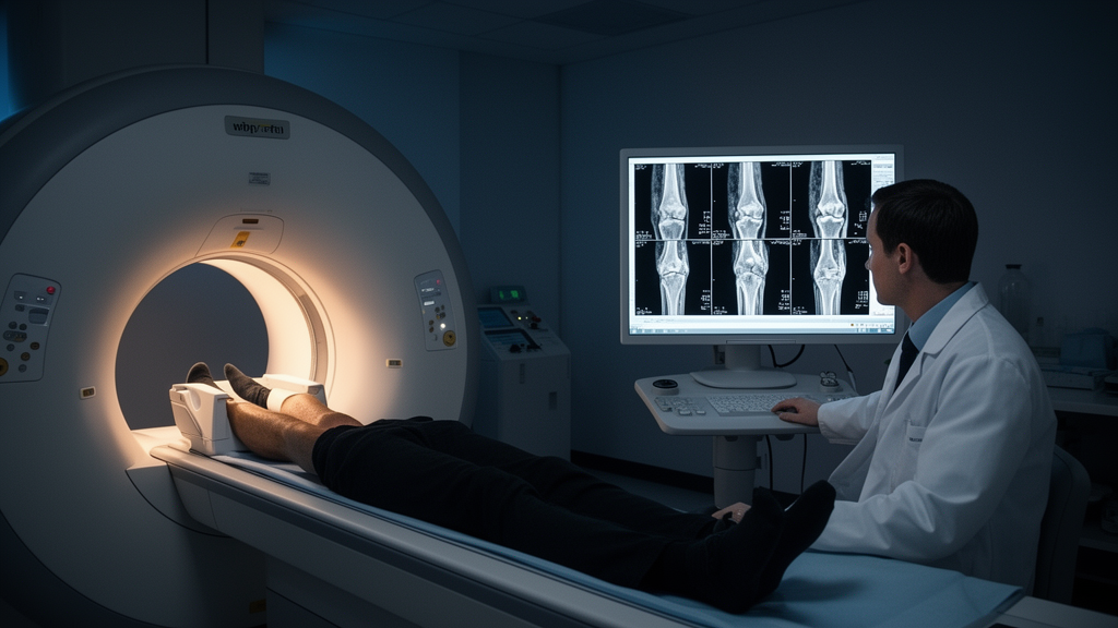
A prospective comparative study published in BMC Musculoskeletal Disorders reports that quantitative T2* mapping values measured on a standard 1.5 T clinical MRI scanner rose steadily across three magnetic resonance imaging-defined categories of the anterior cruciate ligament: normal, partial tear, and complete tear. The median final ACL T2* value was 11.69 milliseconds (interquartile range 11.20–12.04) in the normal ACL group, 14.83 ms (13.46–15.45) in the partial tear group, and 17.52 ms (16.84–22.12) in the complete tear group, with the complete tear group showing the highest values and the normal group the lowest.
Group comparisons using the Kruskal–Wallis test showed a statistically significant difference among the three categories (H = 59.948, p < 0.001), with a large effect size (epsilon-squared = 0.666). Dunn–Bonferroni post hoc analysis showed significant differences between all pairwise group comparisons, meaning normal versus partial tear, partial tear versus complete tear, and normal versus complete tear each differed significantly. Interobserver reliability was excellent, with an overall ICC (2, 1) of 0.998 (95% CI, 0.997–0.998). In adjusted robust multivariable regression with participant-clustered standard errors, ACL category remained significantly associated with the final mean T2* value after adjustment for age, sex, and knee side.
T2* mapping is a quantitative MRI technique that assigns numerical values related to tissue characteristics, potentially offering an objective complement to visual reading. Because the technique can be implemented on clinically available scanners, the findings suggest a measurable quantitative gradient in ACL T2* values that tracks with increasing severity of MRI-defined tear category.
The authors are careful to state the limits of what the study shows. It did not evaluate whether T2* mapping improves the diagnostic accuracy of conventional MRI, provides incremental diagnostic value, or establishes diagnostic thresholds for tear detection. They call for further prospective diagnostic-accuracy studies using an independent reference standard, such as arthroscopy where clinically indicated, before any such role for T2* mapping can be established. As reported, the results describe significant quantitative differences among MRI-defined groups in a comparative observational design; they do not by themselves demonstrate that T2* mapping should change how ACL injuries are diagnosed in practice.
Subject of Research: Quantitative T2* mapping values across MRI-defined normal, partial-tear, and complete-tear anterior cruciate ligament categories
Article Title: T2* mapping values across MRI-defined normal, partial-tear, and complete-tear anterior cruciate ligament categories: a prospective comparative study
Article References: Hari, S., Sekar, K., Lazar, V. R., K, M., Nixon, A., Kumar, E. V., & Aiyappan, S. K. (2026). T2* mapping values across MRI-defined normal, partial-tear, and complete-tear anterior cruciate ligament categories: a prospective comparative study. BMC Musculoskeletal Disorders. https://doi.org/10.1186/s12891-026-10660-z
Image Credits: AI Generated
DOI: 10.1186/s12891-026-10660-z
Keywords: anterior cruciate ligament, T2* mapping, MRI, knee injury, ligament tears, quantitative imaging, musculoskeletal radiology, diagnostic imaging, prospective study, orthopedics, mapping, values
News Source: Ophelia Keating. (October 29, 2026). Quantitative MRI T2* mapping distinguishes normal, partial, and complete ACL tears. Scienmag.



