


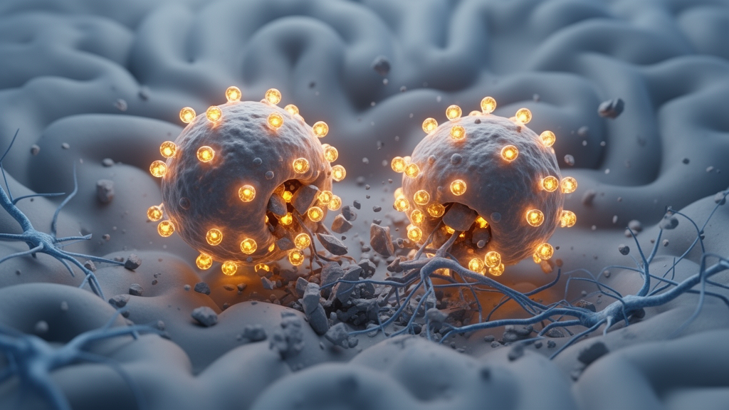
A serotonin receptor found on immune cells that flood the brain after a stroke may determine how well those cells clear away the wreckage of the injury, according to a study published in Nature Neuroscience on 28 October 2026. The work, led by Yueman Zhang, Yonghui Chen and Yue Cheng with senior author Peiying Li, identifies a specific population of monocyte-derived macrophages, or MDMs, that carry the serotonin receptor Htr2b and shows that these cells accumulate in the peri-infarct region of the ischemic brain, where they display enhanced phagocytosis and gene signatures associated with resolving inflammation.
The researchers used single-cell RNA sequencing together with fate mapping in an MDM-specific Ms4a3creERT2-Ai14 mouse line to distinguish infiltrating monocyte-derived macrophages from resident microglia, two brain myeloid cell types that both respond to ischemic injury but whose separate contributions have remained difficult to disentangle. Brain myeloid cells are known to clear debris and shape neuroinflammation and neurological outcomes after stroke, yet the mechanisms governing those functions have been unclear. The sequencing and lineage-tracing approach revealed that the Htr2b-positive MDM subset is a crucial cell population involved in resolving neuroinflammation within the ischemic penumbra, the tissue surrounding the infarct core that is at risk but not yet lost.
Phagocytosis, the process by which macrophages engulf and digest cellular debris, dead tissue and other material, is energetically demanding and depends on a well-functioning lysosomal system, the cell’s network of acidic degradation compartments. The study found that Htr2b activation promotes the nuclear translocation of Tfe3, a transcription factor that serves as a key regulator of lysosomal biogenesis. When Tfe3 moves into the nucleus, it drives the production of lysosomal components, expanding the cell’s digestive capacity. In the Htr2b-positive MDMs, this pathway enhances lysosomal biogenesis and supports the cells’ phagocytic activity, allowing them to internalize and degrade debris more efficiently.
The causal role of this receptor was tested by removing Htr2b specifically from MDMs. These MDM-specific Htr2b-ablated mice developed exacerbated neuroinflammation and ischemic injury after stroke, and their brain macrophages showed impaired debris clearance. Neurological deficits also deteriorated in these animals, as shown in the study’s figures documenting worse ischemic brain injury and poorer functional outcomes compared with controls. The findings indicate that without serotonin signaling through Htr2b, the infiltrating macrophages cannot fully execute the cleanup work that helps limit damage in the penumbra.
The reverse experiment provided the therapeutic counterpart. Pharmacological agonism of Htr2b, delivered intranasally, restored lysosomal biogenesis in brain MDMs and improved post-stroke outcomes, enhancing the phagocytic function of the macrophages and improving neurological outcomes. Direct activation of Tfe3 produced a similar rescue, confirming that the lysosomal regulator sits downstream of the receptor in the pathway. The effect was demonstrated in both murine and human macrophages, where Htr2b signaling promoted efficient phagocytic internalization and intracellular degradation, suggesting the mechanism is not limited to the mouse.
The accumulation of Htr2b-positive MDMs was observed not only in mice but also in the human ischemic brain after stroke, giving the mouse findings a human correlate. Serotonin is conventionally studied as a neurotransmitter acting on neurons, but this work frames it as an extracellular signal that licenses intrinsic phagocytic function in immune cells within the injured brain. The authors describe the Htr2b-positive MDM subset as previously unrecognized, integrating serotonin signaling to support debris clearance and thereby alleviating ischemic brain injury.
The study’s structure, moving from cell identification through mechanism to intervention, links each step to specific evidence. Single-cell RNA sequencing and fate mapping established which cells express Htr2b; imaging and functional assays showed enhanced phagocytosis and inflammation-resolving gene signatures in that subset; genetic ablation demonstrated what happens when the receptor is missing; and pharmacological agonism or Tfe3 activation showed that the pathway can be engaged therapeutically. The figures trace this arc from accumulation in the ischemic brain of mice and humans, through impaired phagocytic function in receptor-deficient mice, to lysosomal biogenesis, Tfe3 nuclear translocation and intranasal agonist delivery.
Because the work was performed in mouse models of acute ischemic stroke with supporting observations in human macrophages and human ischemic tissue, the results establish a mechanism rather than a validated treatment. The intranasal agonist experiments show improved neurological outcomes in the animal model, and the authors present this as a demonstration of a previously unrecognized pathway rather than as a clinical recommendation. Translating the finding would require determining whether Htr2b agonism is safe and effective in human stroke patients, a question the study does not address. Serotonin receptors are expressed in multiple tissues and participate in vascular and immune regulation, so any therapeutic development would need to account for effects beyond the brain’s macrophage population.
What the study does establish is that a specific serotonin receptor on a specific subset of infiltrating macrophages acts as a molecular switch for lysosomal capacity in the injured brain. When that switch is engaged, macrophages clear debris more effectively and the ischemic penumbra fares better; when it is removed, inflammation worsens and injury expands. The identification of Tfe3 as the downstream effector provides a second, potentially druggable node in the same pathway. The work adds to a growing recognition that the immune response to stroke is not uniformly harmful and that specific cell states within the inflammatory infiltrate can actively resolve damage, a distinction that may prove useful for designing therapies that support repair rather than broadly suppress inflammation.
Subject of Research: How serotonin signaling through the Htr2b receptor enhances phagocytosis in brain macrophages after ischemic stroke
Article Title: Serotonin signaling enhances phagocytosis by brain macrophages through Htr2b and alleviates brain injury after stroke
Article References: Zhang, Y., Chen, Y., Cheng, Y., Li, R., Chen, W., Li, Y., Fan, Q., Zhou, Y., Huang, T., Xie, W., Guo, Y., Liesz, A., Wan, J., Ginhoux, F., & Li, P. (2026). Serotonin signaling enhances phagocytosis by brain macrophages through Htr2b and alleviates brain injury after stroke. Nature Neuroscience. https://doi.org/10.1038/s41593-026-02482-y
Image Credits: AI Generated
DOI: 10.1038/s41593-026-02482-y
Keywords: stroke, microglia, macrophages, serotonin, Htr2b, phagocytosis, lysosomes, Tfe3, neuroinflammation, single-cell RNA sequencing, ischemic penumbra, signaling
News Source: Cassandra Pierce. (October 28, 2026). Serotonin Receptor in Brain Macrophages Boosts Debris Clearance After Stroke. Scienmag.



