


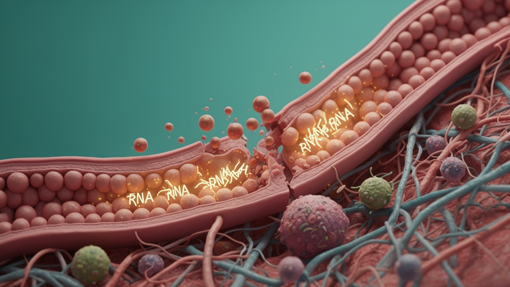
Ductal carcinoma in situ, or DCIS, is a cancerous growth of breast duct cells that may remain indolent or progress to invasive ductal carcinoma. As screening rates increase, the prevalence of DCIS diagnosis has risen, with many women undergoing aggressive treatment with surgery, radiation, and endocrine therapy when diagnosed. Identifying factors that predict progression to invasive disease is therefore critical for improved outcomes, and a new study points to the spatial arrangement of RNAs within the breast tumor microenvironment as a potentially important factor.
The research, published in Breast Cancer Research, examined the tumor microenvironment using breast tissue microarrays representing matched benign (normal adjacent), DCIS, and invasive ductal carcinoma samples. The analysis covered 139 cores, which provided data from 47 unique patients, allowing the team to compare molecular patterns across stages of breast cancer progression within the same study design.
The researchers focused on regulatory RNAs, including both messenger RNAs and long non-coding RNAs. The markers they characterized included Runt-related transcription factor 1 (RUNX1), Runt-related transcription factor 2 (RUNX2), the mitotically activated long non-coding RNA (MANCR), Cluster of Differentiation 90 (CD90), C-X-C motif chemokine 12 (CXCL12), C-X-C chemokine receptor type 6 (CXCR6), and tumor protein 63 (TP63). These molecules were chosen as candidate biomarkers of breast cancer progression.
To visualize where these RNAs sit within tissue, the team used RNAScope fluorescence in situ hybridization, a technique that detects individual RNA molecules in place. In parallel, they used multiplex immunofluorescence to map a panel of stromal marker proteins: Cluster of Differentiation 3 (CD3), Cluster of Differentiation 68 (CD68), Cluster of Differentiation 34 (CD34), alpha smooth muscle actin (aSMA), and TP63. Together, these methods allowed the investigators to examine both the expression levels and the physical distribution of markers in the epithelial and stromal compartments of benign, DCIS, or invasive lesions.
The study identified several temporal and spatial expression signatures of RNAs and/or proteins throughout breast cancer progression. These signatures appeared both in the epithelial and in the stromal compartment of benign, DCIS, or invasive lesions. The findings suggest that the dialogue between tumor epithelium and stroma, a key focus of tumor microenvironment research, can be read not only through which molecules are present but also through where they are located.
A central element of the work was spatial proximity analysis, which assessed the location of markers away from the epithelial boundary or within the myoepithelial layer of the lesions. By quantifying marker positions relative to these structures, the researchers could relate molecular geography to clinical information.
That analysis identified significant associations with clinical parameters, including tumor recurrence status. In their conclusions, the authors report that decreased myoepithelial continuity may be protective against invasive ductal carcinoma recurrence, a finding they suggest could help better patient stratification for response to therapy. The work emphasizes the importance of spatial localization of markers in the breast cancer tumor microenvironment and their importance for clinical outcomes.
Techniques such as RNAScope RNA-FISH and multiplex immunofluorescence preserve positional information, allowing researchers to ask whether the same molecule behaves differently depending on its microanatomical context.
For DCIS specifically, the clinical stakes are considerable. Because many women diagnosed with DCIS receive aggressive treatment, biomarkers that predict which lesions are likely to progress to invasive disease could, if validated, support more tailored management. The authors frame their findings as a step toward identifying such biomarkers, while noting that the value of spatial localization lies in understanding crosstalk between tumor epithelium and stroma and the factors that influence progression of DCIS lesions.
As with any early-stage biomarker study, the results come from a defined sample set of 47 unique patients represented across 139 tissue microarray cores, and the associations reported, including the link between myoepithelial continuity and recurrence, will require further evaluation before they can inform clinical practice. The study was published as open access research, and the authors note that the version shared early is citable and carries a permanent DOI, subject to further edits before the final Version of Record.
Subject of Research: Spatial localization of regulatory RNAs in the breast tumor microenvironment and its association with DCIS progression and recurrence
Article Title: Spatial localization of RNAs within the breast tumor microenvironment may affect progression and recurrence of DCIS
Article References: Lee, K., Bouffard, N. A., Evans, M. F., Balla, A., Weaver, D. L., Sprague, B. L., Stein, J. L., Stein, G. S., Gordon, J. A. R., & Ghule, P. N. (2026). Spatial localization of RNAs within the breast tumor microenvironment may affect progression and recurrence of DCIS. Breast Cancer Research. https://doi.org/10.1186/s13058-026-02402-9
Image Credits: AI Generated
DOI: 10.1186/s13058-026-02402-9
Keywords: DCIS, breast cancer, tumor microenvironment, RNA-FISH, spatial transcriptomics, RUNX1, MANCR, myoepithelial cells, recurrence, biomarkers, tissue microarray, invasive ductal carcinoma
News Source: Nathaniel Bowman. (October 28, 2026). RNA positions in the breast tumor microenvironment linked to DCIS progression and recurrence. Scienmag.



