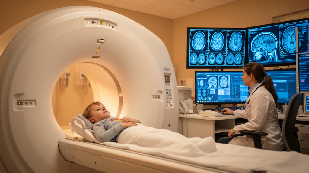
Diffusion spectrum imaging may offer a noninvasive way to grade childhood brain tumors, according to a study published in Cancer Imaging that compared five diffusion-based magnetic resonance imaging models in pediatric patients with intracranial tumors.
Researchers enrolled 44 children with pediatric intracranial tumors, with a mean age of 8.39 years and 18 males among them. All patients underwent diffusion-weighted imaging (DWI) alongside diffusion spectrum imaging approaches: diffusion tensor imaging (DTI), diffusion kurtosis imaging (DKI), mean apparent propagator (MAP) imaging, and neurite orientation dispersion and density imaging (NODDI). The tumors were classified into 23 low-grade and 21 high-grade cases based on histological grading.
The team compared diffusion metrics between the low-grade and high-grade groups using the Mann-Whitney U test, then assessed diagnostic performance with receiver operating characteristic (ROC) analysis and examined relationships with proliferative activity using Spearman correlations.
Several parameters differed significantly between the two grades. Values of MAP_MSD (mean squared diffusion), MAP_QIV (q-space inverse variance), and DWI_ADC (apparent diffusion coefficient) were significantly lower in high-grade tumors, while MAP_NG (non-gaussianity), MAP_NGAx (axial non-gaussianity), MAP_NGRad (radial non-gaussianity), NODDI_ICVF (intracellular volume fraction), and NODDI_ODI (orientation dispersion index) were significantly higher, with p values below 0.05.
All parameters yielded areas under the ROC curve between 0.72 and 0.82. NODDI_ICVF achieved the highest diagnostic performance for distinguishing tumor grades, with an AUC of 0.82 (p < 0.001), and showed significant integrated discrimination improvement over DWI (corrected p < 0.05).
The authors also performed a sensitivity analysis restricted to 27 pediatric gliomas. This analysis indicated the robustness of MAP parameters in pediatric glioma grading, with favorable efficiency reflected in AUCs of 0.75 to 0.82.
Beyond grading, the study examined proliferative activity as measured by the Ki-67 index. MAP, NODDI, and DWI metrics all exhibited significant correlations with this marker of cellular proliferation, suggesting that diffusion-derived measures may reflect biological aggressiveness without requiring tissue sampling.
The authors concluded that MAP and NODDI have diagnostic performance comparable to DWI in grading pediatric intracranial tumors and may provide new approaches for selecting therapeutic strategies. The study is an open-access research article published on 21 October 2026, and the version shared carries a note that it is an accepted manuscript subject to further edits before the final Version of Record.
The work adds to a body of research applying advanced diffusion models—previously explored in meningioma grading, glioma grade evaluation, and the differentiation of high-grade gliomas from brain metastases—to pediatric neuro-oncology.
Subject of Research: Diffusion spectrum imaging for grading and assessing proliferative activity of pediatric intracranial tumors
Article Title: Predictive performance of diffusion spectrum imaging for histological grading and proliferative activity of pediatric intracranial tumors
Article References: Tian, M., Zhang, H., Zhao, S., Lin, Z., Song, Y., Guo, W., Su, Y., Zeng, Z., & She, D. (2026). Predictive performance of diffusion spectrum imaging for histological grading and proliferative activity of pediatric intracranial tumors. Cancer Imaging. https://doi.org/10.1186/s40644-026-01143-4
Image Credits: AI Generated
DOI: 10.1186/s40644-026-01143-4
Keywords: pediatric brain tumors, diffusion spectrum imaging, NODDI, mean apparent propagator, diffusion kurtosis imaging, tumor grading, Ki-67 index, MRI, ROC analysis, diffusion tensor imaging, glioma, Cancer Imaging
News Source: Nathaniel Bowman. (October 28, 2026). Advanced MRI diffusion metrics show promise for grading pediatric brain tumors. Scienmag.