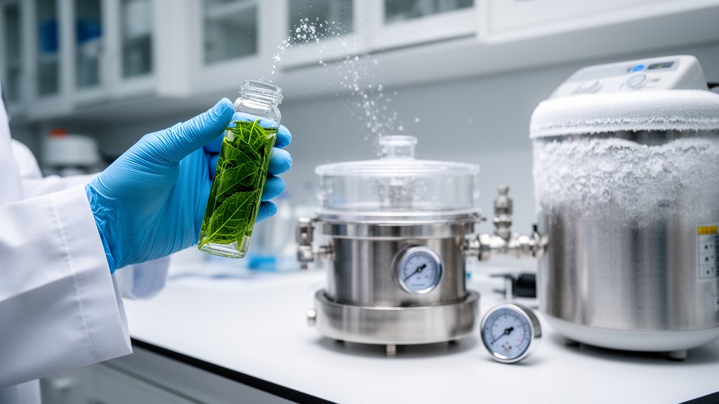
Preparing plant tissue for transmission electron microscopy has long been a technical headache. A new study published in Botanical Studies describes a practical refinement of high-pressure freezing and freeze substitution (HPF-FS) that makes the method work reliably across a range of plant organs, from delicate Arabidopsis root tips to thick orchid petals. The key insight is deceptively simple: before freezing, the tissue should sit in 1-hexadecene inside a vacuum desiccator and be pumped down so that trapped air escapes as visible bubbles. The authors report that the optimal duration of this degassing step scales with the thickness of the sample, giving laboratories a quantitative starting point rather than trial and error.
High-pressure freezing is widely regarded as the gold standard for preparing biological samples for electron microscopy of resin-embedded sections. For freeze-substituted material, the water is later replaced at low temperature by an organic solvent containing stains or crosslinkers, and the sample is then infiltrated with resin and sectioned for imaging or immunolabeling.
Animals and microbes freeze well under these conditions, but plants are another matter. Plant cells are enclosed by walls and, outside meristems, nearly all contain vacuoles of varying size that occupy most of the cell volume. Because vacuoles are essentially dilute solutions of water and soluble substances, the density of cellular solutes is much lower than in animal cells, and freezing efficiency falls as water content rises. Mature leaves, in which vacuoles dominate the cell interior, are therefore notoriously difficult to vitrify. Intercellular spaces add a second problem: in roots they hold water, but in leaves, stems and anthers they hold air, and air is the single greatest obstacle to rapid heat transfer during freezing.
To tackle the air problem, the team, led by Chia-Mei Hsu, Yi-Chia Chou, Hsiao-Jung Yen and Wann-Neng Jane, turned to 1-hexadecene, a hydrophobic paraffin oil with low surface tension that is a common cryoprotectant and filler for plant samples. It permeates into intercellular air spaces and displaces the air, but it cannot do so efficiently while bubbles of gas remain trapped. Earlier attempts with a syringe used to create a micro-vacuum improved results for Arabidopsis anthers but mostly failed for mature leaves, which yielded unclear fine structure. Placing samples in 1-hexadecene inside a vacuum desiccator connected to a vacuum pump proved far more effective: as the pressure dropped to 2 × 10⁻² torr, numerous air bubbles were released from the tissue.
The researchers then systematically varied the degassing time, testing 3, 5, 8, 10, 12 and 15 minutes, and froze the samples in a Leica EM PACT2 high-pressure freezer at 2020 to 2050 bar with a freezing rate exceeding 10,000 degrees Fahrenheit per second. Frozen samples were transferred to a Leica AFS2 freeze-substitution unit at −160 °C, held for five minutes, then warmed to −90 °C. Substitution solutions included 2% osmium tetroxide in acetone or 0.1% uranyl acetate plus 1% osmium tetroxide in acetone for morphology, and 0.1% uranyl acetate plus 0.2% glutaraldehyde in ethanol for immunolabeling. The substitution schedule ran for several days, stepping gradually from −90 °C up to 20 °C, followed by resin infiltration and polymerization in Spurr’s resin, EMBed 812 or LR Gold depending on the application.
The results demonstrate how the optimal degassing time tracks tissue thickness. Arabidopsis root tips, 1.2 millimeters long and taken from 14-day seedlings, needed no degassing at all, because the intercellular spaces in the root tip region are minimal and filled with water rather than air. Arabidopsis anthers at the bicellular pollen stage were well preserved after just three minutes of pumping, showing intact anther walls with epidermis, endothecium, middle layer and tapetum, pollen cells with lipid bodies surrounding the generative cell, and tapetal cells containing elaioplasts and tapetosomes whose stacked ER cristae were clearly resolved.
Leaves demanded longer treatment in proportion to their thickness. Mature leaves of Brachypodium distachyon, a C3 grass roughly 80 micrometers thick, gave their best results after six minutes of degassing, revealing mesophyll cells with chloroplasts whose thylakoid membranes and stacked grana were sharply defined, along with mitochondria bearing well-developed cristae. Axonopus compressus, a C4 grass with leaves about 100 micrometers thick, required eight minutes; its dimorphic chloroplasts were preserved with starch grains abundant in bundle sheath cells and fewer in mesophyll cells, and plasmodesmata were visible in the walls between bundle sheath cells and between bundle sheath and mesophyll. Plantago asiatica, a campus weed in Taiwan with leaves of 180 to 200 micrometers, needed ten minutes, and its phloem, with sieve tube members retaining only mitochondria and companion cells packed with mitochondria, showed crisp ultrastructure throughout.
The most demanding tissue was the petal of the scented orchid Phalaenopsis bellina, which exceeds 600 micrometers in thickness at the basal region on the first day of blooming. Twelve minutes of degassing produced the best outcome. Petal sections showed epidermal and cortical cells dominated by large vacuoles, epidermal cells with prominent nuclei and electron-transparent channels in their outer walls, cortical cells with rough ER arranged parallel to the cell walls, abundant exocytic vesicles between the plasma membrane and cell wall, and plasmodesmata traversing the walls. The authors note that in earlier attempts without degassing, cortical cells ruptured and organelles were unrecognizable, and even with degassing by syringe the intracellular contents were destroyed by ice crystals. Only the vacuum-desiccator approach preserved both cell integrity and fine structure.
Across all tissues, the authors report a positive correlation between sample thickness and the optimal degassing duration, and they argue that the true variable is the volume of air held in intercellular spaces, which corresponds to thickness. Because the vacuum level can be controlled, the method provides a quantitative basis for planning freeze runs rather than relying on intuition. They also observed that the negatively stained thylakoid membranes reported for Arabidopsis leaves under standard freeze-substitution protocols did not appear in their preparations, suggesting their schedule avoided that artifact. A practical caveat concerns 1-hexadecene itself: because it only dissolves when warmed above −68 °C, frozen 1-hexadecene coating the sample must be removed before substitution can proceed at −85 or −90 °C, and if the oil has seeped into intercellular spaces, the time below −70 °C and the warming ramp must be extended.
The study positions HPF-FS as a near-complete replacement for chemical fixation in plant electron microscopy, whether for morphological observation, immunolocalization or tomography, while acknowledging that achieving good results is harder than with traditional methods. By documenting working parameters for roots, anthers, leaves of three species with contrasting anatomy and a thick orchid petal, and by showing that degassing time can be predicted from tissue thickness, the work offers plant cell biologists a reproducible recipe for capturing cellular architecture in a state far closer to life than chemical fixation allows.
Subject of Research: A vacuum degassing method to improve high-pressure freezing and freeze substitution of plant tissues for transmission electron microscopy
Article Title: Sample preparation of some plant tissues for transmission electron microscopy using high pressure freezing and freeze substitution
Article References: Hsu, C.-M., Chou, Y.-C., Yen, H.-J., & Jane, W.-N. (2026). Sample preparation of some plant tissues for transmission electron microscopy using high pressure freezing and freeze substitution. Botanical Studies, 67(1), Article 13. https://doi.org/10.1186/s40529-026-00499-x
Image Credits: AI Generated
DOI: 10.1186/s40529-026-00499-x
Keywords: high-pressure freezing, freeze substitution, transmission electron microscopy, plant tissue, 1-hexadecene, degassing, Arabidopsis thaliana, ultrastructure, vacuoles, intercellular air spaces, Phalaenopsis bellina, cryofixation
News Source: Drew Townsend. (October 27, 2026). Vacuum degassing unlocks high-pressure freezing for tough plant tissues. Scienmag.