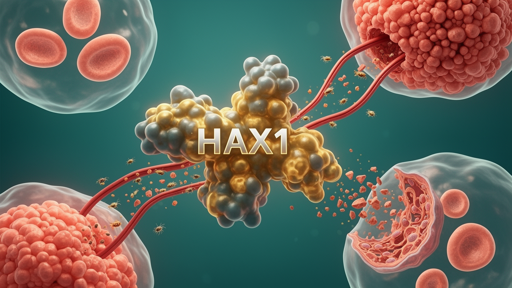
A 2026 review concludes that HCLS1-associated protein X-1, or HAX1, is not a conventional enzyme but a flexible molecular scaffold whose abundance, location and binding partners can help tumors survive stress, multiply, invade tissue and resist therapy. Across breast, gastric, colorectal, lung, liver, nasopharyngeal and other cancers, elevated HAX1 has been associated with more aggressive clinicopathological features and poorer survival in retrospective clinical studies. Experimental evidence further links HAX1 to mitochondrial and endoplasmic-reticulum stress protection, proliferative signaling, cytoskeletal remodeling, hypoxic adaptation, angiogenesis and multiple forms of treatment resistance. The findings nominate HAX1 as a prognostic biomarker and a possible therapeutic target, but the evidence remains predominantly based on laboratory models and retrospective patient data rather than successful clinical intervention.
HAX1 was first identified as a binding partner of hematopoietic cell-specific protein 1, from which its name derives. The canonical human isoform contains 279 amino acids and is encoded by a gene on chromosome 1q21.3. It is found mainly in mitochondria, but also occurs in the endoplasmic reticulum, cytoplasm, nuclear membrane, vesicles and RNA granules. Its distribution is not static: in migrating cells, it can accumulate near the plasma membrane at the leading edge of lamellipodia. HAX1 is broadly but unevenly expressed across human tissues, with relatively high RNA levels in skeletal muscle and lower levels in lung. Because its unbound structure is predominantly disordered, HAX1 can acquire local secondary structures when it binds lipid membranes, while alternative splicing produces isoforms with different sequences and intracellular distributions. This flexible architecture supports its role as an intrinsically disordered scaffold that assembles protein complexes rather than catalyzing a single defined reaction.
The protein’s cancer-related functions depend heavily on context-specific interactions. HAX1 binds the beta-6 cytoplasmic tail of integrin alpha-v-beta-6 and regulates its clathrin-mediated endocytosis, a process associated with tumor migration and invasion. It also interacts with HS1 and the actin-binding protein cortactin, linking it to cytoskeletal remodeling. Other partners connect HAX1 to the endoplasmic-reticulum calcium pump SERCA2 and the mitochondrial chaperone CLPB, extending its influence over calcium homeostasis and mitochondrial proteostasis. Although HAX1 lacks a canonical RNA-binding domain, it binds the 3-prime untranslated regions of vimentin and DNA polymerase beta messenger RNAs and associates with additional RNAs involved in ribosomal RNA processing, ribosome assembly, cytoskeletal organization and protein translation. These broad interactions explain how one scaffold can influence gene expression, translation, cell structure and stress adaptation at the same time.
Noncoding RNAs help determine how much HAX1 is produced. MicroRNAs generally bind the 3-prime untranslated region of HAX1 messenger RNA and reduce its expression, whereas long noncoding and circular RNAs can sequester those microRNAs and relieve repression. In hepatocellular carcinoma, miR-125a-5p was reported to directly target HAX1, with its suppression associated with reduced tumor-cell growth, migration and epithelial-mesenchymal transition in the experimental models examined. The long noncoding RNA CYTOR can attenuate miR-125a-5p-mediated repression, preserving HAX1 and reducing mitochondrial caspase-9 activation and apoptosis. A comparable axis operates in esophageal squamous cell carcinoma, where circGFPT1 sequesters miR-142-5p and increases HAX1 expression. Conversely, miR-125b directly targets HAX1 in breast cancer cells; restoring its repression of HAX1 increased sensitivity to doxorubicin. Nasopharyngeal carcinoma involves an additional positive feedback loop in which HAX1 binds and stabilizes IGF2BP1 messenger RNA, while the N6-methyladenosine reader IGF2BP1 stabilizes HAX1 messenger RNA in return.
Ubiquitination controls a different layer of HAX1 regulation by determining its stability, degradation or condensation. A PEST sequence in HAX1 promotes lysine-48-linked polyubiquitination and proteasomal degradation. In mantle cell lymphoma, protein kinase C delta phosphorylates HAX1 and the E3 ubiquitin ligase substrate receptor FBXO25, promoting FBXO25-dependent HAX1 degradation; disrupting this axis allows HAX1 to accumulate and weakens apoptosis. In colorectal cancer, the translation-initiation factor EIF3H acts as a deubiquitinase that removes beta-TrCP-mediated polyubiquitin chains from HAX1. Stabilized HAX1 then promotes assembly of the RAF1–MEK1–ERK1 complex, linking the protein’s abundance to proliferative signaling. RPS4X can also suppress degradation by disrupting the Skp1–Cullin1–F-box ubiquitin ligase complex. A different ubiquitin linkage changes HAX1’s physical state rather than simply its lifetime: under energy stress, TRIM23 catalyzes lysine-63-linked polyubiquitination, inducing liquid-liquid phase separation and processing-body assembly. These condensed RNA granules suppress global protein synthesis and support colorectal cancer-cell proliferation during nutrient deprivation, while high cytoplasmic TRIM23, HAX1 expression and HAX1 ubiquitination were associated with poor outcomes in patient samples.
Many of HAX1’s tumor-promoting effects arise from its protection against programmed cell death. Its interaction with the non-receptor tyrosine kinase c-Abl supports reactive oxygen species clearance under stress, while binding to the stress-response protein AATF, also called Che-1, influences p53 activity and mTOR signaling. Oxidative stress strengthens the HAX1–AATF interaction in MCF7 breast cancer cells. HAX1 also regulates SERCA2 and cellular calcium stores while suppressing mitochondrial caspase-9 activation, limiting cell death caused by oxidative stress, disturbed calcium balance and mitochondrial apoptosis. Gene-silencing experiments in uveal melanoma and cervical carcinoma support this protective role. In non-small cell lung cancer, HAX1 sustains AKT/mTOR survival signaling and MDM2-mediated suppression of p53, so HAX1 depletion can restore p53 activity and induce apoptosis; a similar AKT1-dependent survival function has been reported in glioblastoma. The review also identifies HAX1-dependent focal-adhesion turnover, Rac1–cortactin complex formation and NF-kappa-B-dependent epithelial-mesenchymal transition as mechanisms through which it can facilitate migration and invasion.
HAX1 can also help malignant cells adapt to a low-oxygen environment and recruit blood vessels. In glioma models, hypoxia-inducible factor 1 alpha increases HAX1 expression, after which HAX1 promotes AKT-dependent phosphorylation of the dynamin-related protein Drp1 and mitochondrial fission, allowing cells to maintain growth and invasive behavior under hypoxia. At the vascular level, tumor-derived extracellular vesicles can transfer HAX1 to endothelial cells. There, HAX1 increases translation of integrin beta-6 and activates focal adhesion kinase, promoting endothelial migration and tube formation; in the preclinical models studied, HAX1-rich vesicles also increased metastasis in vivo. These observations are consistent with the wider proposal that HAX1 supports tumor progression through multiple connected mechanisms, but they do not establish that HAX1 is uniformly required in every cancer or that inhibiting it would produce a clinically meaningful effect in patients.
Clinical studies support HAX1’s potential as a biomarker, although most of the evidence is retrospective. HAX1 positivity was detected in 74.4% of nasopharyngeal carcinoma samples compared with 20.9% of non-tumor nasopharyngeal tissues, while moderate or strong staining occurred in 61.7% of cutaneous squamous cell carcinomas and 3.3% of normal skin samples. HAX1 was also enriched in serum extracellular vesicles from patients with nasopharyngeal carcinoma relative to healthy donors, raising the possibility of investigation in liquid biopsy studies. High HAX1 expression was independently associated with adverse prognosis in gastric cancer, colorectal cancer, nasopharyngeal carcinoma, laryngeal carcinoma and glioma. In multiple myeloma, high HAX1 expression was reported to be associated with poor survival, although the review does not describe that association as independently prognostic. In gastric cancer, three-year overall survival was 41.5% in the high-expression group and 64.6% in the low-expression group. HAX1 expression correlated with tumor classification, nodal metastasis, stage, size or histological grade in several cancers. Its subcellular location may also matter: in breast cancer, cytoplasmic HAX1 independently predicted distant recurrence, whereas nuclear HAX1 did not. In resected early-stage non-small cell lung cancer, the rs11265425 G allele increased HAX1 promoter activity and was associated with shorter disease-free survival, principally in adenocarcinoma.
Therapeutic strategies emerging from the reviewed evidence focus less on inhibiting an HAX1 enzyme than on disrupting selected interactions or preventing abnormal stabilization and condensation. A cell-penetrating HAX1 peptide competitively displaced the protein from integrin beta-6 in oral squamous cell carcinoma cells, blocking alpha-v-beta-6 internalization and reducing motility and invasion. In ovarian carcinoma models, expression of a competitive HAX1 fragment called HAX D4 prevented Rac1–cortactin complex formation and reduced lysophosphatidic acid-induced migration by more than 75%. Perturbing the HAX1–IQGAP1 interaction can also impair focal-adhesion turnover and directional migration. Other experiments target the regulators of HAX1: EIF3H depletion in colorectal cancer restores HAX1 ubiquitination and weakens RAF1–MEK1–ERK1 assembly, and blocking TRIM23-mediated lysine-63 ubiquitination impairs processing-body formation and tumor growth. HAX1 depletion has also enhanced cisplatin activity in nasopharyngeal carcinoma models, sensitized hepatocellular carcinoma cells to a ferroptosis inducer, and reversed the camptothecin-resistant phenotype in SW480 colorectal cancer cells. These are preclinical combination findings, not evidence of clinical efficacy.
The principal obstacle is that HAX1 also supports normal physiology. Germline loss-of-function variants cause severe congenital neutropenia, indicating that systemic and indiscriminate inhibition could damage essential hematopoietic functions. The review therefore argues that selective strategies should distinguish tumor-specific dependencies from HAX1 activity required by healthy blood and immune cells. Existing clinical evidence is also dominated by retrospective expression analyses, often without prospective validation or demonstration that HAX1 adds prognostic information beyond established clinicopathological factors. Future studies need to test whether expression, subcellular localization, molecular modification state and binding-partner profiles can identify patients most likely to benefit. A safer development path would compare HAX1-directed interventions in tumor models with their effects on normal hematopoietic cells, while prospective studies assess whether HAX1 measurements predict recurrence, treatment response or survival. The current evidence establishes HAX1 as a versatile scaffold and promising marker, but not yet as a proven cancer treatment.
Subject of Research: Review of HAX1 molecular regulation, prognostic value and therapeutic vulnerabilities across cancers
Article Title: HAX1 in cancer: from molecular scaffolding to therapeutic targeting
Article References: Sun, T., Xu, D., Zhou, T., Luo, F., & Zhang, Y. (2026). HAX1 in cancer: from molecular scaffolding to therapeutic targeting. Frontiers in Oncology, 16, Article 2000988. https://doi.org/10.3389/fonc.2026.2000988
Image Credits: AI Generated
DOI: 10.3389/fonc.2026.2000988
Keywords: HAX1, cancer biology, molecular scaffold, tumor progression, prognostic biomarker, treatment resistance, ubiquitination, noncoding RNA, hypoxic adaptation, targeted therapy, cancer, molecular
News Source: Nathaniel Bowman. (October 26, 2026). HAX1 review highlights context-dependent roles in cancer progression. Scienmag.