Calcific aortic valve disease affects more than a quarter of people over the age of 65, yet no drug has been shown to halt or reverse its progression. A new study now points to an unexpected mechanism behind the disease's spread through valve tissue: calcifying cells package mitochondrial material into tiny membrane-bound vesicles and release them into their surroundings, where they push neighboring cells toward bone-like mineralization. The research, conducted on human valve tissue, primary human valve cells, and mouse models, identifies a specific molecular pathway—centered on the secreted protein Gas6—that controls the final step by which these vesicles detach from their parent mitochondria.
The study focused on human valvular interstitial cells, the resident cell population of the aortic valve and central players in calcific aortic valve disease pathogenesis. A clinically striking feature of valvular calcification is its spatial pattern: mineral deposits begin at discrete foci and extend outward into adjacent tissue. This point-to-surface progression raised the possibility that calcifying cells actively influence their neighbors through extracellular mediators rather than simply degenerating in place.
The researchers set out to identify what those mediators might be and how their release is regulated. Their attention turned to mitochondrial-derived vesicles, membranous particles roughly 70 to 150 nanometers in diameter that bud off from mitochondria and can carry damaged mitochondrial proteins, lipids, and mitochondrial DNA. These vesicles are heterogeneous, and their cargo composition reflects the nature and severity of the cellular stress that produced them.
Vesicles carrying exclusively outer mitochondrial membrane markers or exclusively matrix components are generated under basal conditions or mild oxidative stress. Vesicles harboring both outer membrane and matrix constituents arise under more severe metabolic dysfunction and have been linked to disease processes in prior work. To test whether such vesicles are released from calcifying human valves, the team developed an ex vivo hemodynamic simulation system.
Aortic valve leaflets were obtained from 12 patients aged 60 to 73 years—six undergoing surgical valve replacement for severe calcific aortic valve disease and six age- and sex-matched patients undergoing cardiac surgery for pure aortic regurgitation without valve calcification. After six hours of pulsatile perfusion at physiological frequency, the circulating medium was collected and extracellular vesicles were isolated by differential ultracentrifugation. Immunoblotting confirmed the presence of classical vesicle markers alongside the mitochondrial outer membrane protein TOM20, while the cytoplasmic marker beta-actin was undetected, indicating clean vesicle isolation.
Nanoparticle tracking showed peak vesicle diameters of approximately 180 nanometers. Nano-flow cytometry then revealed that calcified valve leaflets released significantly higher levels of TOM20-positive mitochondrial-derived vesicles than non-calcified controls. The critical functional question was whether these vesicles could transmit calcifying instructions.
Using TOM20-conjugated magnetic beads to enrich the mitochondrial-derived vesicle population, the researchers applied preparations from calcified or non-calcified valve tissue to naïve primary human valvular interstitial cells maintained in control medium. Alizarin Red S staining showed that vesicles from calcified valves induced substantial calcium deposition in recipient cells, while those from non-calcified valves produced minimal mineralization. Vesicles from calcified tissue also increased protein levels of the osteogenic markers RUNX2, osteopontin, and BMP2 in recipient cells.
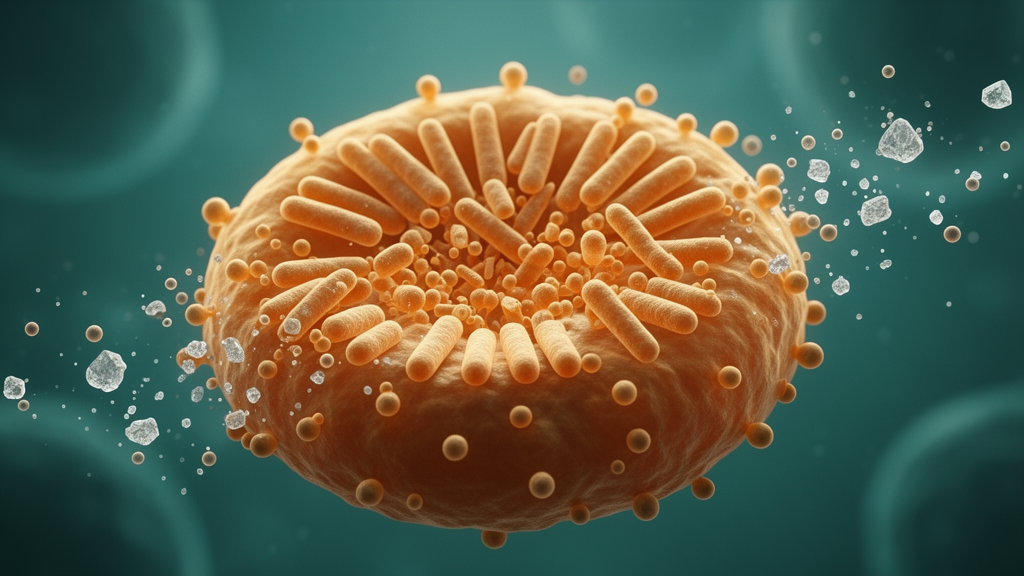
A parallel experiment using purified human valvular interstitial cells cultured in pro-osteogenic medium recapitulated these findings: cells under osteogenic conditions released more vesicles in a dose-dependent manner, and vesicles from those cells triggered mineral deposition and osteogenic marker expression in naïve recipients. High-resolution imaging revealed the physical platform from which these vesicles emerge. Under osteogenic conditions, human valvular interstitial cells exhibited TOM20-positive, PDH-positive vesicular buds contiguous with hollow, donut-shaped mitochondrial structures—closed, toroidal organelles that had not previously been visualized in human tissue or primary human valvular cells.
Three-dimensional confocal reconstruction, STED super-resolution microscopy, and transmission electron microscopy all confirmed this morphology, and electron microscopy of calcified human valve tissue showed donut-shaped mitochondrial profiles absent from non-calcified controls. Live-cell imaging captured the temporal sequence: mitochondria curl and self-fuse into donut shapes, then bud off vesicles that detach and enter the extracellular space. To identify molecular drivers of this process, the team integrated previously published proteomic and transcriptomic datasets from human calcific and non-calcific valve tissue, identifying 36 candidates upregulated at both transcript and protein levels.
Among them was Gas6, growth arrest-specific 6, recently annotated as a candidate senoprotein in a human lifespan proteomic atlas. Independent immunoblotting and immunofluorescence confirmed higher Gas6 protein expression in calcific valve tissue, paralleling RUNX2 upregulation, with spatial overlap between the two proteins. The calcific tissue and osteogenically treated cells also showed senescence-associated features—elevated p16INK4A and p21, positive senescence-associated beta-galactosidase staining in calcified regions—consistent with the age-associated nature of the disease.
Functional experiments established Gas6 as a driver of both calcification and vesicle release. Silencing Gas6 with siRNA reduced RUNX2, osteopontin, and BMP2 protein levels under osteogenic conditions and diminished calcium nodule formation; adenoviral overexpression produced the opposite effects. Critically, live-cell time-lapse imaging revealed that Gas6 knockdown did not prevent mitochondria from curling into donut-shaped precursors—the proportion of such structures remained comparable between control and Gas6-silenced cells, and the mitochondrial fission gene signature was unaltered.
Instead, Gas6-deficient cells displayed a striking defect at the final scission step: vesicles formed but failed to detach, remaining tethered to the parent mitochondrion in a stalled budding state. This finding localized Gas6's role precisely: it does not initiate vesicle formation but enables the terminal membrane fission event that releases vesicles into the extracellular space. Downstream signaling analysis identified the pathway through which Gas6 drives this terminal scission.
Gas6 knockdown reduced calcium influx, diminished CaMKII phosphorylation, and lowered the activation of DRP1, the dynamin-related protein that executes mitochondrial fission. These results establish a linear signaling axis: Gas6 promotes calcium entry, which activates CaMKII, which in turn phosphorylates and activates DRP1 to drive the terminal membrane scission that releases pro-calcific vesicles from donut-shaped mitochondria. In vivo experiments confirmed the pathway's physiological relevance.
The findings collectively demonstrate that Gas6-dependent terminal scission of donut-shaped mitochondria releases pro-calcific vesicles that propagate calcification through valve tissue, positioning this pathway as a potential target for future therapeutic investigation in calcific aortic valve disease.
Subject of Research: Gas6-dependent release of pro-calcific mitochondrial-derived vesicles in calcific aortic valve disease
Article Title: The Senokine Gas6 Promotes Aortic Valve Calcification Through CaMKII–DRP1–Mediated Terminal Scission of Mitochondrial‐Derived Vesicles
Article References: Zhang, Y., Zhang, J., Xu, Y., Lei, W., Xu, F., Li, J., Wei, Z., Su, B., Liu, S., Zhou, J., Li, H., Ran, J., Li, W., Tuerdi, T., Wang, X., Fang, Z., Huang, F., & Chen, M. (2026). The Senokine Gas6 Promotes Aortic Valve Calcification Through CaMKII–DRP1–Mediated Terminal Scission of Mitochondrial‐Derived Vesicles. Aging Cell, 25(11), Article e70758. https://doi.org/10.1111/acel.70758
Image Credits: AI Generated
DOI: 10.1111/acel.70758
Keywords: calcific aortic valve disease, mitochondrial-derived vesicles, Gas6, CaMKII, DRP1, valvular interstitial cells, mitochondrial fission, senescence, senoproteins, aortic stenosis, vascular calcification, ex vivo perfusion
News Source: Beatrice Stafford. (October 24, 2026). Aging Valve Cells Release Pro-Calcific Vesicles Through a Gas6-Driven Mitochondrial Scission Pathway. Scienmag.