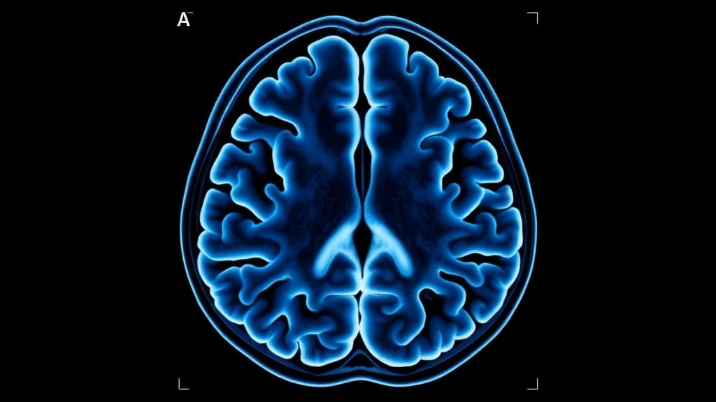
Researchers in Bangladesh have published the first comprehensive set of normative values for brain ventricular measurements derived from computed tomography scans of healthy Bangladeshi adults, filling a conspicuous gap in a clinical field long dominated by Western reference data. The retrospective cross-sectional study, conducted across Chattogram Medical College Hospital and two private diagnostic centers from January to December 2023, analyzed brain CT images from 314 apparently healthy individuals, 161 men and 153 women, aged over 18 years. The team measured eight widely used ventricular indices and found that most showed no significant difference between the sexes, while ventricular size increased measurably with advancing age, particularly after the age of 60 and particularly in men. The findings give clinicians in Bangladesh a locally derived baseline for distinguishing normal anatomical variation from pathological ventricular enlargement, a distinction that underpins the diagnosis of hydrocephalus, cerebral atrophy, and neurodegenerative disease.
The ventricular system is a series of interconnected fluid-filled cavities deep within the brain: two lateral ventricles nestled in the cerebral hemispheres, a slit-like third ventricle in the midline between the halves of the diencephalon, and a fourth ventricle lying between the brainstem and the cerebellum. Together they produce, circulate, and remove cerebrospinal fluid, the medium that cushions the brain mechanically and clears metabolic waste. When these spaces enlarge or change shape, the change can signal hydrocephalus, traumatic brain injury, or neurodegenerative disorders such as Alzheimer’s and Parkinson’s disease. The difficulty for clinicians is that ventricular enlargement also occurs naturally with aging, so without population-specific baseline measurements, separating normal variation from pathology becomes a matter of guesswork. Linear indices measured on axial CT scans, which benefit from high inter-observer reliability, offer a standardized way to make that call.
The study team included only Bangladeshi individuals by birth whose CT scans had been reported as normal by an experienced radiologist, meaning the absence of trauma, hydrocephalus, space-occupying lesions, hemorrhage, or other structural abnormalities. Anyone with a history of head injury, cerebral ischemia, hemorrhagic infarction, previous intracranial surgery, alcoholism, or drug abuse was excluded. The sample size of 320 was calculated from Indian normative data on the Evans index, with a standard error of 0.003, and 314 participants ultimately met the criteria. All scans were acquired on a Siemens Somatom 128-slice multi-detector CT scanner, with axial sections taken from the skull base to the vertex at 5 mm slice thickness, referenced to the cantho-meatal line and acquired at 15 to 20 degrees above it. Measurements were performed in DICOM software, and any scan flagged as abnormal was removed from the analysis.
Each index captures a different aspect of ventricular morphometry. The Evans index is the ratio of the transverse diameter of the anterior horns of the lateral ventricles to the internal diameter of the skull, measured at the level of the frontal horns. The bifrontal index divides the maximum bifrontal diameter, a line connecting the two anterior corners of the frontal horns, by the brain width at the same level. The bicaudate index, measured at the level of the caudate nuclei heads, divides the minimum lateral ventricular width by the inner skull width, making it a sensitive marker of subcortical atrophy. The Huckman index sums the transverse diameter of the anterior horns and the minimum lateral ventricular width. Third ventricular width was measured at the widest point of the third ventricle on the thalamic level, and the third ventricular index divides that width by the transverse brain diameter. The antero-posterior and transverse diameters of the fourth ventricle were taken at its widest visible axial section.
The overall results establish the normative values at the heart of the study. The mean third ventricular width was 3.74 mm, with a standard deviation of 1.46, and the third ventricular index averaged 0.032. The antero-posterior diameter of the fourth ventricle averaged 6.519 mm and its transverse diameter 11.873 mm. The bifrontal index recorded a mean of 0.312, the Evans index 0.253, and the bicaudate index 0.122. Huckman’s number averaged 44.778 mm. The distribution of Evans index values followed a unimodal pattern, with the vast majority of measurements falling below the conventional hydrocephalus diagnostic threshold of 0.3, exactly what one would expect in a genuinely healthy sample. The average participant age was 38.8 years, with men slightly older at 39.8 years than women at 37.7 years, a difference that was not statistically significant.
Sex comparisons revealed a consistent but selective pattern. Men had significantly wider third ventricles, 3.92 mm versus 3.55 mm in women, a difference with a p-value of 0.011. The transverse diameter of the fourth ventricle was notably larger in men, 12.228 mm against 11.499 mm, with a p-value below 0.001. The bicaudate index showed a small but significant male predominance, 0.126 versus 0.118, and Huckman’s number was significantly elevated in men, 46.427 versus 43.042, again with a p-value below 0.001. By contrast, the third ventricular index, the Evans index, the bifrontal index, the ventricular index, and the antero-posterior diameter of the fourth ventricle showed no statistically significant sex differences. The authors suggest that this pattern of minimal sex-based variation could allow a unified reference range to be applied in routine neuroimaging assessment within the Bangladeshi population, simplifying clinical decision-making.
Age told a more dramatic story. Post-hoc pairwise comparisons using Tukey’s test showed that the majority of ventricular indices increased at later ages, particularly after the age of 60. In the 61-to-70 age group, men exhibited a third ventricular index of 0.046 and a fourth ventricular transverse diameter of 12.98 mm, while their Huckman number reached 50.58, compared with 46.94 in women of the same age band. In the over-70 group, men recorded a Huckman index of 48.61 against 47.17 in women. Crucially, the authors note, most of these age-related values remained within normal limits, supporting the interpretation that such enlargement reflects the normal aging process rather than a marker of pathology. This has a direct clinical consequence: in elderly patients, Evans index values approaching the conventional 0.3 threshold may represent physiological aging, whereas values exceeding it, especially when accompanied by gait disturbance, cognitive decline, or urinary incontinence, should raise strong suspicion for hydrocephalus or cerebral atrophy and warrant further diagnostic work-up.
The comparison with international data affirms both the local and global utility of the measurements. The Bangladeshi Evans index mean of 0.253 sits almost exactly on the Nepalese figure of 0.251 and within the 0.252-to-0.255 range reported in Nigerian and Ghanaian studies, while Indian studies reported slightly higher values of 0.26 to 0.27. The third ventricular index closely matched the Indian value of 0.034, and the fourth ventricular diameters aligned with South Indian and Nigerian data, suggesting relatively similar ventricular dimensions across South Asian and African populations. The bifrontal index mean of 0.312 mirrored Indian normative values of 0.31 to 0.33. The consistency, the authors argue, supports the validity of the widely accepted 0.3 Evans index threshold for hydrocephalus even in this population, and contrasts with some Indian studies that reported significant sex-based variability in indices where the Bangladeshi sample found none, a discrepancy attributed to ethnic and methodological differences in skull and brain size.
The study is not without constraints, and the authors are candid about them. The retrospective design may introduce selection bias, since only individuals who underwent neuroimaging for minor indications were included. CT-based linear measurements cannot capture the full complexity of three-dimensional ventricular volume change, and the single-center setting restricts generalizability across the broader Bangladeshi population. The lack of MRI validation, a modality offering superior soft-tissue and volumetric resolution, and the absence of inter-rater reliability assessment further limit the findings. The authors call for multi-center studies across Bangladesh, longitudinal assessments to track ventricular change over time, and direct comparison of linear CT indices with volumetric MRI datasets to refine local reference standards.
The practical payoff is nonetheless substantial. Locally derived normative values enhance diagnostic confidence when evaluating suspected hydrocephalus or cerebral atrophy and reduce inappropriate reliance on Western thresholds, which could misinterpret ventricular size given documented population differences in cranial morphometry, such as the smaller cranial breadths reported in African and Asian populations compared with Europeans. The consistency of the Bangladeshi data with international figures also demonstrates the reproducibility of standard CT-based measurement techniques in resource-limited settings. Beyond the clinic, the authors point to a forward-looking application: indices such as the Evans and bicaudate indices are measurable with automated tools, making this normative dataset a potential training and validation resource for artificial intelligence-based diagnostic systems. Early identification of subtle ventricular changes through these indices, the study concludes, can facilitate timely clinical decision-making and improve outcomes for patients with neurological disease.
Subject of Research: Neuroanatomy and neuroimaging
Article Title: Normative values of ventricular indices of brain using computed tomography scans in Bangladeshi adults: a cross-sectional study
Article References: Basak, D., Roy, S., Tasmia, K., Yesmin, N., Shuvo, A. U., & Hasan, M. J. (2026). Normative values of ventricular indices of brain using computed tomography scans in Bangladeshi adults: a cross-sectional study. Heliyon, 12(16), Article e45618. https://doi.org/10.1016/j.heliyon.2026.e45618
Image Credits: AI Generated
DOI: 10.1016/j.heliyon.2026.e45618
Keywords: neuroimaging, computed tomography, ventricular indices, Bangladesh, normative values, hydrocephalus, brain anatomy, cerebrospinal fluid, Evans index, cross-sectional study, Normative, values
News Source: Cassandra Pierce. (October 24, 2026). CT Scans Reveal First Brain Ventricular Norms for Healthy Bangladeshi Adults. Scienmag.