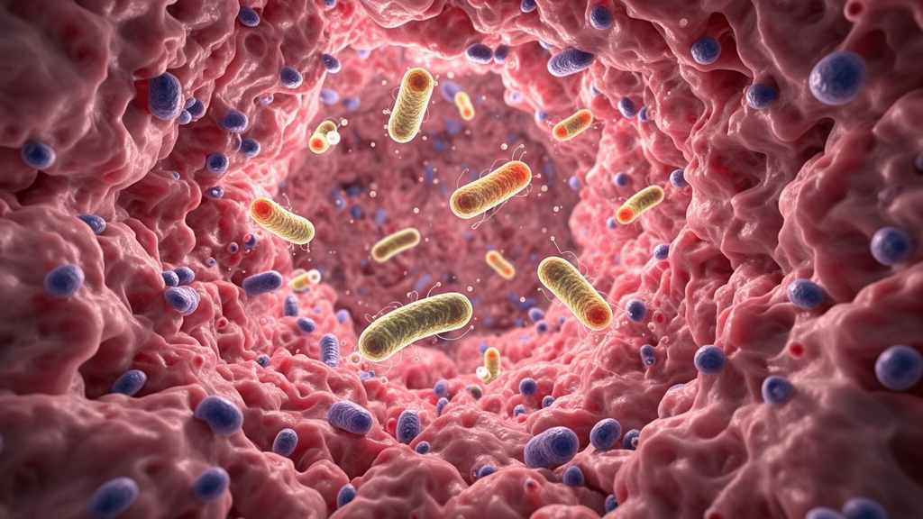
Deep inside gliomas, the most common malignant tumors of the central nervous system, researchers have found communities of bacteria whose composition appears to track how dangerous the tumor is and how long the patient is likely to live. A retrospective study of 107 treatment-naïve glioma patients, published in Frontiers in Cellular and Infection Microbiology, used 16S rRNA gene sequencing of archived tumor tissue to profile the microbes residing within the tumors, then linked those microbial profiles to World Health Organization grade and overall survival. The work challenges the long-held view of the brain as a sterile fortress and adds a surprising new dimension to an already complicated picture of glioma biology.
The clinical stakes are high. Gliomas are graded from I to IV, and patients with grade IV tumors typically survive less than two years despite aggressive multimodal therapy. Modern classification, codified in the fifth edition of the WHO Classification of Tumors of the Central Nervous System, incorporates molecular features such as IDH mutation status, MGMT promoter methylation, CDKN2A/B deletion, TERT promoter mutation, EGFR alterations, and the characteristic +7/−10 copy-number changes. Yet outcomes remain stubbornly heterogeneous even among patients with similar molecular subtypes, implying that other biological determinants remain to be discovered. The tumor microbiome, sometimes called the second genome, has emerged in recent years as one candidate dimension, with microbes documented inside multiple solid tumors where they appear to modulate immune responses, metabolic pathways, and therapeutic sensitivity.
The research team, led by investigators at the Second Affiliated Hospital of Chongqing Medical University and the National University of Singapore, extracted DNA from formalin-fixed paraffin-embedded tumor specimens using the CTAB method, amplified the V4 hypervariable region of the bacterial 16S rRNA gene, and sequenced the libraries on an Illumina NovaSeq 6000. Because intratumoral samples carry very low microbial biomass, contamination control was a central concern. Paraffin-only blanks and no-tissue extraction controls were processed in parallel, and any amplicon sequence variants that dominated the negative controls or showed background-level distribution patterns were removed before analysis, along with low-prevalence and extremely low-abundance variants that could reflect reagent contamination or index hopping.
Across the 107 specimens, the team detected 61 microbial taxa at the phylum level. Proteobacteria, Firmicutes, Actinobacteriota, and Bacteroidetes dominated, and core taxa such as Proteobacteria and Firmicutes appeared in more than 90 percent of samples. Rare phyla accounted for the largest share of the diversity, comprising 33 of the 61 identified phyla, or 54.1 percent, underscoring substantial heterogeneity within the glioma microenvironment. At lower taxonomic levels, the team noted enrichment of genera including Escherichia–Shigella and Streptococcus, which they suggest could reflect altered blood–brain barrier permeability or abnormal microbial infiltration into the tumor niche.
To connect microbes with outcome, the researchers split the cohort using an optimal survival cut-off of 664 days, which separated 37 favorable-prognosis patients from 70 poor-prognosis patients. Microbial diversity, measured by both Shannon and Simpson indices, was significantly higher in the poor-prognosis group. Differential abundance analysis then revealed contrasting signatures: taxa enriched in the favorable group included Firmicutes, Clostridia, Lachnospirales, Lachnospiraceae, and several short-chain fatty acid–producing bacteria, while the poor-prognosis group was characterized by Porphyromonas gingivalis, Escherichia–Shigella, Streptococcus sobrinus, Vibrio species, and Alphaproteobacteria. Notably, however, principal coordinate analysis based on Bray–Curtis dissimilarity showed overlapping profiles between the two groups, and PERMANOVA did not detect a statistically significant overall compositional difference, with PERMDISP also indicating unequal dispersion, which limits interpretation of the apparent clustering as a genuine difference in community location.
The team then built predictive models. Random forest screening selected the top 30 survival-associated features, which fed into LASSO regression. In the final model, Firmicutes emerged as the strongest positively associated microbial predictor, with a coefficient of −0.2462, followed by Clostridia at −0.1300, while age, sex, Galbibacter marinus, Alphaproteobacteria, and Porphyromonas gingivalis were retained as risk-associated variables. Time-dependent ROC analysis produced apparent AUCs of 0.78, 0.86, and 0.90 at one, two, and three years, and calibration curves showed good agreement between predicted and observed survival. After bootstrap optimism correction with 1,000 resamples, the corrected AUCs were 0.7005, 0.8723, and 0.9591.
Grade-related microbial patterns proved even more informative. High-grade tumors harbored more diverse communities, with median Shannon index values of approximately 5.90 versus 5.25 in low-grade tumors, and taxa enriched in high-grade samples included Galbibacter marinus, Porphyromonas gingivalis, Peptostreptococcus anaerobius, Fusobacteriota, Vibrio species, Enterobacterales, Escherichia–Shigella, Acinetobacter species, and Moraxellaceae. Low-grade samples were instead characterized by Firmicutes-related taxa such as Clostridia, Lachnospirales, Lachnospiraceae, Bacillales, Bacillaceae, and Burkholderiales. A WHO grade–informed model combining these microbial features with clinical parameters achieved apparent AUCs of 0.79, 0.86, and 0.91 at one, two, and three years, and optimism-corrected AUCs of 0.7105, 0.8597, and 0.8828. In exploratory analyses of the 37 patients with lipid data, microbial features outlasted serum metabolic parameters as predictors, with Clostridia and Firmicutes retained in the final grade-informed model.
Functional inference through PICRUSt2 added a metabolic layer to the story. Comparing favorable and poor-prognosis groups, the researchers found enrichment of pathways related to energy metabolism, nucleotide biosynthesis, and amino acid metabolism in the poor-prognosis tumors, including the tricarboxylic acid cycle, de novo purine biosynthesis, de novo pyrimidine biosynthesis, tRNA charging, GDP-mannose biosynthesis, and S-adenosylmethionine synthesis. Comparing low- and high-grade tumors, de novo purine biosynthesis showed the most pronounced difference, with pyrimidine salvage, de novo pyrimidine biosynthesis, inosine 5′-phosphate degradation, and glycine heme biosynthesis also enriched in high-grade disease. Protein–protein interaction network analysis identified 33 core genes centered on enzymes of mitochondrial energy metabolism such as SDHA, OGDH, and FH, nucleotide synthesis such as IMPDH1, GART, ATIC, and DTYMK, and amino acid metabolism and translation such as GDH1, MARS1, and WARS1. Enzymes like IMPDH1 and GART have previously been linked to cancer cell growth and therapeutic vulnerability in other contexts.
The authors are careful to frame these findings as associations rather than demonstrations of causation. Detection of microbial DNA in fixed tissue does not establish that viable bacteria are present or reveal their spatial localization within the tumor, and the PICRUSt2 functional profiles are computationally inferred rather than measured. The molecular marker analysis suffered from limited power, since only 24 of the 107 samples had complete molecular data, and markers including IDH mutation, CDKN2A deletion, 1p/19q co-deletion, MGMT methylation, EGFR alterations, and +7/−10 changes did not reach statistical significance in that subset. Information on extent of resection and performance status was unavailable for some patients, and the possibility that part of the microbial signal reflects reagent- or environment-derived contamination in these low-biomass samples cannot be excluded, despite the stringent controls.
Even with those caveats, the study suggests that intratumoral microbiota profiling could one day complement histopathology, molecular markers, and neuroimaging in glioma risk stratification. Prior work has shown that Porphyromonas gingivalis can promote tumor cell proliferation through gingipain-mediated activation of MAPK signaling and sustained inflammation, while short-chain fatty acids such as butyrate have been reported to suppress glioma cell proliferation and enhance radiosensitivity in experimental models, raising the possibility of microbiome-informed therapeutic strategies ranging from dietary interventions to targeted microbial modulation. Such approaches would need to be applied cautiously, since indiscriminate microbial manipulation could disrupt beneficial communities. For now, the Chongqing and Singapore team’s message is measured: microbial signatures inside gliomas are associated with malignancy grade, predicted metabolic remodeling, and patient survival, and independent prospective validation with spatial microbial confirmation and multi-omics integration will be essential before these observations translate into clinical practice.
Subject of Research: Associations between intratumoral microbiota, glioma malignancy grade, and patient survival based on 16S rRNA sequencing of 107 tumor specimens
Article Title: Intratumoral microbiota stratifies glioma malignancy and patient survival through metabolic associations
Article References: Yao, Y., Yang, Y., Song, J., Wu, L., Xu, M., Liu, H., Liou, Y.-C., & Xia, L. (2026). Intratumoral microbiota stratifies glioma malignancy and patient survival through metabolic associations. Frontiers in Cellular and Infection Microbiology, 16, Article 1940097. https://doi.org/10.3389/fcimb.2026.1940097
Image Credits: AI Generated
DOI: 10.3389/fcimb.2026.1940097
Keywords: glioma, intratumoral microbiota, 16S rRNA sequencing, tumor microbiome, WHO grading, overall survival, prognostic modeling, nucleotide biosynthesis, Firmicutes, Clostridia, Porphyromonas gingivalis, glioblastoma
News Source: Cassandra Pierce. (October 24, 2026). Bacteria Living Inside Brain Tumors Track How Aggressive Gliomas Are. Scienmag.