A surgical technique that grafts a small island of gum tissue onto an exposed tooth root has delivered measurable improvements in both function and appearance for orthodontic patients with fragile gums, according to a new clinical study from Shanghai. Researchers at Shanghai Ninth People’s Hospital, Shanghai Jiao Tong University School of Medicine, report that their modified connective tissue graft, known as the epithelium island (EI) technique, simultaneously widened keratinized gum tissue, thickened the gingiva, and reduced visible root exposure in fifteen patients with thin periodontal phenotypes. The findings, published in Frontiers in Oral Health, were verified not only with traditional periodontal probing but also with a fully digital workflow combining intra-oral scanning, cone beam computed tomography, and an objective color-difference formula.
The clinical problem the technique addresses is a familiar one in orthodontics. Patients with thin periodontal phenotypes, meaning thin gingiva overlying the tooth roots, are more likely to develop gingival recession than those with thick phenotypes, and untreated recessions in thin phenotypes tend to progress even when oral hygiene is good. Thin-phenotype patients also tend to have insufficient keratinized tissue, the tough, pale band of gum that resists mechanical stress. Clinically, at least 2 mm of keratinized tissue width is recommended to avoid periodontal problems, including the progression of recession. For patients lacking sufficient gingiva or keratinized tissue, periodontal phenotype modification therapies aim to increase the width and thickness of these tissues, reducing the risk of adverse outcomes such as clinical attachment loss while supporting esthetic root coverage.
The established gold standard for treating gingival recession is the coronally advanced flap combined with a subepithelial connective tissue graft, the so-called bilaminar technique. However, recent systematic reviews and meta-analyses cited by the authors indicate that this gold standard shows lower predictability in sites with inadequate baseline keratinized tissue width or deep recession defects, where complete root coverage rates may fall from the typical 70 to 85 percent to around 40 to 55 percent. For patients without sufficient keratinized tissue, the traditional solution has been a two-step approach first described by Bernimoulin and colleagues in 1975: a free gingival graft is placed apical to the exposed root to augment keratinized gingiva, and roughly two months later a coronally advanced flap is performed to cover the root. Zucchelli and De Sanctis later proposed an improved two-step variant combining free gingival grafts with coronally advanced or laterally positioned flaps.
While two-step techniques can be valid in challenging cases, they carry inevitable drawbacks: the process is time-consuming, and the secondary operation increases patient pain. Color mismatch with the adjacent gingiva remains a major concern for free gingival grafts, which often produce conspicuous white scarring. Modified single-stage approaches such as the partly epithelialized free gingival graft and the gingival unit graft have attempted to combine keratinized tissue augmentation with root coverage, but the authors note these still face limitations, including inadequate keratinized tissue augmentation or unclear gingival thickness gains, and comprehensive digital evaluations of their outcomes have not been fully reported. The epithelium island technique was designed to close this gap by merging the keratinized tissue augmentation of a free gingival graft with the root coverage potential of a connective tissue graft in one procedure.
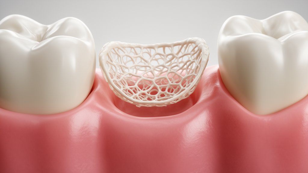
The surgical design is the heart of the innovation. A horizontal incision is made at the mucogingival junction of the affected tooth, and a split-flap is prepared apical to the incision. The coronal area of the incision is then de-epithelialized. The graft itself is harvested from the palate in two connected portions: a 1 mm thick connective tissue component harvested using the L technique, and a 2 mm thick epithelium-containing component harvested in the manner of an epithelium-connective tissue graft, with a length ratio between the two parts of 1 to 1.5. The epithelial portion is placed over the affected tooth and sutured with horizontal cross mattress sutures, while the connective tissue component is secured beneath the split-flap of adjacent teeth with interrupted sutures. Unlike a conventional free gingival graft, the connective tissue component of the EI graft is covered by a gingival flap, ensuring superior blood supply and promoting graft survival. The epithelial portion, supported by the underlying connective tissue, resembles an island, which gives the technique its name.
The study enrolled fifteen patients, nine female and six male, with a mean age of 27.60 years, each with one affected tooth, for fifteen teeth in total. All had Cairo RT-2 localized gingival recession with keratinized tissue width below 1 mm and a thin periodontal phenotype confirmed by the periodontal probe transparency test. Eleven affected teeth were in the mandible and four in the maxilla. Patients were excluded if they had uncontrolled systemic diseases, were pregnant, or smoked. All participants completed initial periodontal therapy until full-mouth plaque and bleeding scores fell below 20 percent, and clinical parameters were recorded before surgery and at six months postoperatively.
The digital evaluation pipeline was central to the study’s precision. Intra-oral scans were captured with the 3Shape TRIOS 4 scanner, which has a stated accuracy of plus or minus 10 micrometers, extending to the base of the vestibular sulcus and palatal mucosa. CBCT images with a voxel size of 0.125 mm were acquired to evaluate alveolar bone volume and fused with the scan data using 3Shape Implant Studio software. Registration proceeded in stages: at least three shared anatomical landmarks on dental hard tissues were manually marked for initial registration, fine alignment followed using occlusal and labial feature areas, and multi-planar rotation verified the fit between roots and alveolar bone. Pre- and post-surgery scans were then superimposed to measure keratinized tissue width and gingival thickness at 0, 1, and 3 mm apical to the gingival margin, while the root exposure area was calculated automatically from a closed curved region of interest bounded by the gingival margin, the most apical extension of the recession, and the interdental papilla midlines.
The results were substantial. Recession depth fell from 3.00 plus or minus 0.76 mm at baseline to 0.90 plus or minus 0.43 mm at six months, and recession width dropped from 6.07 plus or minus 0.80 mm to 3.73 plus or minus 0.80 mm, both statistically significant. Keratinized tissue width rose from a baseline of 0.60 plus or minus 0.12 mm to 3.45 plus or minus 0.31 mm at six months, an absolute gain of 2.85 plus or minus 0.28 mm. Gingival thickness of affected teeth increased by 0.73, 1.09, and 1.03 mm at 0, 1, and 3 mm apical to the gingival margin respectively, with the largest increment at 1 mm apical. Adjacent teeth showed similar gains, reflecting the graft’s connective tissue extension beneath neighboring flaps. The root exposure area shrank from 5.85 plus or minus 0.97 square millimeters to 0.76 plus or minus 0.42 square millimeters, a reduction of 5.10 plus or minus 1.07 square millimeters. Probing depth, plaque scores, and bleeding scores remained stable throughout, indicating good oral hygiene and no periodontal deterioration.
Esthetic outcomes were assessed objectively by a blinded periodontist using self-developed GAES software, which calculates gingival color differences with the CIEDE2000 formula under standardized D65 illumination at 6500 K, following ISO/TR 28642. Color coordinates of affected teeth showed no significant differences from adjacent teeth at any time point. The color difference values at two weeks, when sutures were removed, were close to baseline, but by six months they had fallen significantly, by 0.865 at the free gingival margin and 0.845 at the middle zone of keratinized gingiva. All values remained at or below 2.7, the threshold for an acceptable color match under the ISO/CIE standard, and the six-month values of 1.22 and 1.34 approached the 1.1 threshold for an excellent match, confirming that the grafted tissue blended with the surrounding gingiva over time.
The authors acknowledge important limitations. The sample was small at fifteen patients, follow-up lasted only six months, and the absence of a control group prevents direct comparison with free gingival grafts or the bilaminar technique. They call for larger cohorts, follow-up of one to two years to assess long-term stability, and randomized controlled designs. They also see a digital future for this kind of assessment, noting that the fusion of intra-oral scans and CBCT data improves the accuracy and reliability of periodontal soft tissue evaluation, and that artificial intelligence could eventually automate tissue segmentation, registration, and measurement of pre- and postoperative periodontal parameters. Within those limits, the study supports the epithelium island technique as a predictable single-stage option for orthodontic patients with thin gums and insufficient keratinized tissue, offering simultaneous functional reconstruction and esthetic improvement where conventional approaches have struggled.
Subject of Research: A modified connective tissue graft technique for treating gingival recession in orthodontic patients with thin periodontal phenotypes
Article Title: Digitally clinical and esthetic assessment on a modified connective tissue graft (epithelium island) technique
Article References: Liao, Y., Hu, H., Li, H., Chen, H., Sun, W., Dong, J., & Song, Z. (2026). Digitally clinical and esthetic assessment on a modified connective tissue graft (epithelium island) technique. Frontiers in Oral Health, 7, Article 1889034. https://doi.org/10.3389/froh.2026.1889034
Image Credits: AI Generated
DOI: 10.3389/froh.2026.1889034
Keywords: gingival recession, connective tissue graft, epithelium island technique, periodontal phenotype, orthodontics, keratinized tissue, intra-oral scanning, CBCT, CIEDE2000, esthetic dentistry, periodontology, root coverage
News Source: Ophelia Keating. (October 23, 2026). Epithelium Island Graft Technique Restores Receding Gums in Orthodontic Patients. Scienmag.