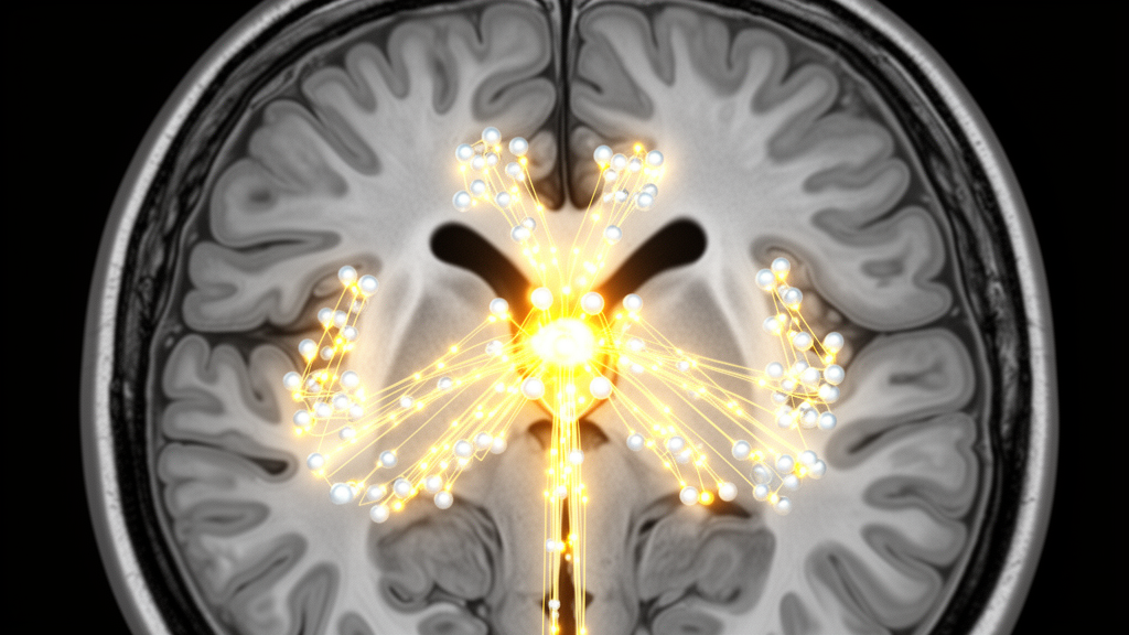
Every time you hold your eyes steady on a target, your brainstem must convert a brief burst of movement commands into a sustained signal that keeps the eyes locked in place. Two small nuclei perform this mathematical trick: the prepositus hypoglossi nucleus (PHN) for horizontal gaze and the interstitial nucleus of Cajal (INC) for vertical gaze. A new study from Nara Medical University, published in Frontiers in Cellular Neuroscience, reports that neurons in the INC respond to acetylcholine with inward currents carried by both nicotinic and muscarinic receptors, and that the cholinergic fibers reaching this vertical gaze integrator originate almost entirely from the caudal magnocellular region of the medial vestibular nucleus (MVeMC), not from the mesopontine tegmentum that had long been assumed to supply such inputs to gaze-holding circuitry.
The research team, Taketoshi Sugimura and Yasuhiko Saito of the Department of Neurophysiology, addressed three questions. Do INC neurons show current responses to acetylcholine at all? Are those responses mediated mainly by nicotinic acetylcholine receptors (nAChRs), as previously shown for the PHN, or does the balance differ by cell type? And where do the cholinergic inputs to the INC actually come from? The answers matter because previous work had established that cholinergic signaling helps the horizontal integrator sustain the firing needed for gaze holding, yet no study had ever reported a direct cholinergic effect on INC neurons, even though fluorescently labeled cholinergic axons had been seen threading through the nucleus in transgenic rats.
For the physiological experiments, the researchers used brainstem slices from VGAT-Venus transgenic rats, in which inhibitory neurons fluoresce because they express vesicular GABA transporter-linked Venus protein. Neurons lacking this fluorescence were classified as putative glutamatergic. Using whole-cell patch-clamp recordings from 38 INC neurons held at minus 75 millivolts, the team applied acetylcholine locally through a pressurized puff pipette at 30-second intervals. Every single INC neuron tested produced an inward current in response to acetylcholine, indicating that cholinergic transmission exerts an excitatory influence on the vertical gaze integrator at the membrane current level.
The magnitude of those responses, however, was notably smaller than in the PHN. When current amplitude and charge transfer were normalized to membrane capacitance, INC neurons averaged 1.44 plus or minus 1.64 picoamperes per picofarad and 0.58 plus or minus 0.52 picocoulombs per picofarad, compared with 4.86 plus or minus 3.14 pA/pF and 1.29 plus or minus 0.91 pC/pF in PHN neurons reanalyzed from earlier work, differences that were highly significant. The comparison was validated by recording a fresh dataset of PHN neurons under the current setup, which showed no significant differences from the previously published values for either glutamatergic or inhibitory cells. Even after excluding the PHN’s own cholinergic neurons from the comparison, INC responses remained smaller, a pattern the authors suggest may reflect a lower density of acetylcholine receptors in the vertical integrator.
Pharmacological dissection revealed a cell-type-specific receptor dependency that distinguishes the two integrators. By bath-applying 5 micromolar atropine to block muscarinic receptors and 10 micromolar mecamylamine to block nicotinic receptors, the researchers found that in glutamatergic INC neurons the nicotinic contribution, 59.3 plus or minus 20.0 percent of current and 56.9 plus or minus 23.3 percent of charge, was not significantly different from the muscarinic contribution. In inhibitory INC neurons, by contrast, nicotinic receptors carried the larger share, 62.0 plus or minus 17.1 percent of current and 62.0 plus or minus 22.0 percent of charge, both significantly exceeding the muscarinic fractions. This contrasts with the PHN, where nicotinic receptors predominated in both glutamatergic cells, at 63.4 plus or minus 13.2 percent of current, and inhibitory cells, at 70.6 plus or minus 18.8 percent. The two integrators, in other words, share a modulatory source but not a uniform receptor strategy.
To trace the anatomical origin of the cholinergic fibers, the team turned to ChAT-tdTomato transgenic rats, in which every cholinergic neuron expresses red fluorescent protein. They injected a retrograde adeno-associated virus vector, AAVrg-hSyn-EGFP, unilaterally into the INC of three animals and waited three weeks for transport. Although a few green-labeled neurons appeared in the pedunculopontine and laterodorsal tegmental nuclei, the classic cholinergic hubs of the mesopontine tegmentum, not a single neuron in those regions was double-labeled with both EGFP and tdTomato. Instead, double-labeled cholinergic neurons clustered densely in the caudal medial vestibular nucleus, specifically its magnocellular region, with the highest density between bregma minus 12.24 and minus 12.60 millimeters, at roughly 14 neurons per three sections on the injected side and 15 on the opposite side.
The parallel tracing experiment in the PHN produced an equally striking result. Despite long-standing assumptions that the mesopontine tegmentum is the primary cholinergic source for the horizontal integrator, the researchers again found no double-labeled cholinergic neurons in either the pedunculopontine or laterodorsal tegmental nuclei. The retrogradely labeled cholinergic neurons instead appeared in the caudal medial vestibular nucleus and, notably, in the caudal PHN itself, meaning some cholinergic neurons within the horizontal integrator project back onto it. The authors caution that their focal injections did not cover the entire PHN, so arousal-related cholinergic inputs from the tegmentum cannot be ruled out, but the data clearly redirect attention to the lower brainstem as the dominant cholinergic supply for both integrators.
This shared origin carries functional weight. The rostral medial vestibular nucleus receives primary vestibular afferents and drives the vestibulo-ocular reflex directly, whereas the caudal region, particularly the magnocellular compartment, is reciprocally connected with the vestibulocerebellum, a loop crucial for calibrating gaze stabilization. The authors propose that cholinergic projections from the MVeMC may convey vestibular signals processed through this cerebellar circuit to sustain the firing of both integrators, providing an anatomical substrate for coordinating horizontal and vertical gaze holding in three dimensions. The finding also echoes the team’s earlier observation that the INC and PHN rely on different excitatory receptor mechanisms within their local circuits, predominantly NMDA receptors in the INC and calcium-permeable AMPA receptors in the PHN, suggesting that distinct receptor dependencies characterize both the core networks and the modulatory systems of each integrator.
The authors are careful about what the study does and does not establish. The puff application of 1 millimolar acetylcholine is substantially diluted before reaching the recorded cell and does not reproduce the spatial and temporal dynamics of endogenous cholinergic transmission; the protocol was designed instead to measure receptor-mediated response capability under standardized, saturating conditions near the soma and proximal dendrites, where voltage clamp is most reliable. Slice recordings also omit the background synaptic and neuromodulatory milieu of the intact brain, and the study did not test whether the acetylcholine-induced inward currents enhance firing rate or persistent spiking, so the link between cholinergic signaling and actual gaze-holding performance remains to be demonstrated directly. Viral tropism and injection spread were addressed as potential biases, with the authors arguing that the robust labeling in the caudal MVeMC makes the absence of tegmental labeling unlikely to be a technical artifact.
Within those boundaries, the study delivers a clear reorganization of how neuroscientists should picture the cholinergic control of gaze. Both the vertical and horizontal neural integrators draw their principal cholinergic innervation from the same caudal vestibular source, yet each deploys its own mixture of nicotinic and muscarinic receptors, weighted differently across glutamatergic and inhibitory cell types, to translate that common input into the sustained activity that holds the eyes still. The work provides a foundation for understanding how vestibular, cerebellar, and cholinergic signals converge to stabilize gaze, and it reframes the mesopontine tegmentum’s role in this circuit as an open question rather than a settled assumption.
Subject of Research: Cholinergic input origins and receptor-mediated current responses in rat interstitial nucleus of Cajal neurons
Article Title: Cholinergic input origins and current response properties in rat interstitial nucleus of Cajal neurons
Article References: Sugimura, T., & Saito, Y. (2026). Cholinergic input origins and current response properties in rat interstitial nucleus of Cajal neurons. Frontiers in Cellular Neuroscience, 20, Article 1925102. https://doi.org/10.3389/fncel.2026.1925102
Image Credits: AI Generated
DOI: 10.3389/fncel.2026.1925102
Keywords: interstitial nucleus of Cajal, prepositus hypoglossi nucleus, gaze holding, neural integrator, acetylcholine, nicotinic receptors, muscarinic receptors, medial vestibular nucleus, retrograde tracing, whole-cell recording, brainstem, oculomotor control
News Source: Drew Townsend. (October 23, 2026). Rat Brainstem Study Traces Acetylcholine Signals That Steady Gaze to a Single Vestibular Source. Scienmag.