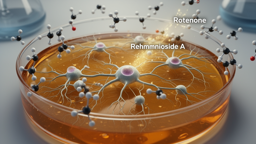
Rehmannioside A, an iridoid glycoside isolated from the traditional Chinese medicinal plant Rehmanniae Radix, protected dopaminergic function and reduced oxidative stress in laboratory models of Parkinson’s disease, according to a study published in Frontiers in Neurology. The experiments combined rotenone-challenged PC12 cells with a rat model in which animals received daily rotenone injections for 28 days. Across both systems, Rehmannioside A improved measures of cell viability, motor performance, neuronal integrity and dopamine-related biochemistry. Its effects were associated with increased phosphorylation of PI3K and Akt and increased expression of tyrosine hydroxylase, the rate-limiting enzyme in dopamine synthesis. The researchers concluded that the compound’s protective effects are at least partly dependent on PI3K/Akt signaling, although the experiments do not establish that the compound can treat Parkinson’s disease in people or prove that it penetrates the human brain.
The cellular experiments used PC12 cells, an undifferentiated rat pheochromocytoma line widely employed as a screening platform for intracellular signaling. Exposure to 1 micromolar rotenone, a mitochondrial complex I inhibitor, significantly reduced cell viability after 24 hours. Pretreatment with Rehmannioside A restored viability to varying degrees across a concentration range of 10–200 micromolar, leading the team to select 25, 50 and 100 micromolar for subsequent analyses. In these groups, rotenone increased malondialdehyde, a terminal product of lipid peroxidation, while reducing glutathione content and superoxide dismutase activity. Rehmannioside A shifted all three indicators toward the untreated control condition in a dose-dependent pattern. Western blotting also showed that rotenone lowered phosphorylated PI3K, phosphorylated Akt and tyrosine hydroxylase, whereas Rehmannioside A attenuated those reductions.
To test whether the cellular response depended on PI3K/Akt signaling, the researchers added LY294002, a pharmacological inhibitor of PI3K. In rotenone-challenged PC12 cells, LY294002 partially reversed Rehmannioside A’s antioxidant effects: it restored higher malondialdehyde and lower glutathione and superoxide dismutase activity relative to the Rehmannioside A-only condition. The inhibitor also weakened the compound-associated increases in phosphorylated PI3K, phosphorylated Akt and tyrosine hydroxylase. LY294002 alone aggravated rotenone-induced oxidative stress. These results support a contribution from PI3K/Akt signaling, but they do not definitively establish causality because LY294002 is not fully specific to PI3K and the investigators did not use genetic knockdown of PI3K or Akt. The authors note that targeted knockdown would provide a more specific test of the pathway’s role.
In the animal experiment, 8- to 10-week-old male Sprague–Dawley rats weighing 260 ± 20 grams were randomly assigned to six groups of six animals. Besides control and rotenone-model groups, the study included three Rehmannioside A doses—20, 40 and 80 milligrams per kilogram of body weight—and a positive-control group given levodopa plus carbidopa. Except for controls, all animals received subcutaneous rotenone at 2 milligrams per kilogram daily for 28 consecutive days. Rehmannioside A was administered orally once daily, beginning on the same day as rotenone exposure and continuing throughout model induction. Because treatment began before or alongside toxin exposure rather than after established degeneration, the design primarily tests a neuroprotective or concomitant effect and does not demonstrate the ability to reverse an already established Parkinson’s-like condition.
Rotenone-modeled rats began losing weight from day 7 and developed the behavioral abnormalities used to confirm model induction, including bradykinesia, unstable gait and postural imbalance. In the rotarod test, control animals remained on the rotating apparatus longer than model animals, whereas all three Rehmannioside A doses and levodopa plus carbidopa significantly extended latency to fall relative to untreated model rats. The open-field test produced a parallel pattern: rotenone reduced rearing episodes and total distance traveled, while each Rehmannioside A dose and the positive control increased both measures compared with the model group. The authors described the rescue of weight loss and motor performance as attenuation rather than complete normalization, and the article did not provide the underlying numerical values and error estimates in its text.
Anatomical analysis indicated that rotenone also damaged neurons in the substantia nigra pars compacta. Hematoxylin and eosin staining revealed marked neuronal injury and pyknosis in model animals, changes that were visibly less pronounced after Rehmannioside A and levodopa plus carbidopa treatment. Immunohistochemistry showed that the percentage of tyrosine hydroxylase-positive cells fell significantly after rotenone exposure and rose dose-dependently with Rehmannioside A. Brain-tissue Western blots produced a related pattern: rotenone lowered phosphorylated PI3K, phosphorylated Akt and tyrosine hydroxylase protein levels, while Rehmannioside A restored the signaling proteins in a dose-dependent manner. Tyrosine hydroxylase upregulation was strongest at the highest dose of 80 milligrams per kilogram. High-dose Rehmannioside A and levodopa plus carbidopa had comparable effects on these endpoints, but the researchers cautioned that this does not imply an identical mechanism, because levodopa primarily replaces deficient dopamine rather than directly activating PI3K/Akt phosphorylation.
The biochemical findings connected the behavioral and tissue observations to the nigrostriatal dopamine system. Rotenone significantly decreased striatal dopamine while increasing its metabolites 3,4-dihydroxyphenylacetic acid, also known as DOPAC, and homovanillic acid, or HVA. Rehmannioside A increased dopamine in a dose-dependent manner and reduced both metabolites relative to model animals. The pattern was consistent with improved dopaminergic function after rotenone injury, although the researchers did not calculate the combined DOPAC-plus-HVA-to-dopamine turnover ratio and therefore could not quantify that aspect of metabolic change. In parallel, Rehmannioside A reduced the elevated striatal malondialdehyde associated with the model and increased the depleted glutathione and superoxide dismutase activity. Together, these measures indicated less oxidative stress and better preservation of dopamine metabolism rather than a complete return of every biomarker to control levels.
The study places Rehmannioside A within a broader effort to find compounds that regulate PI3K/Akt, a signaling axis involved in cell survival, metabolism and antioxidant defenses. Previous research had reported protective effects of Rehmannioside A in experimental vascular dementia, spinal cord injury and ischemic cognitive impairment, with antioxidant, anti-inflammatory and PI3K/Akt-related mechanisms implicated. The new work extends those observations to rotenone-based Parkinson’s models and links reduced oxidative stress with preservation of tyrosine hydroxylase, dopamine-related measures and motor behavior. Tyrosine hydroxylase is relevant because reduced expression is an early feature of dopaminergic degeneration, but the authors stress that staining for this enzyme alone cannot distinguish the survival of dopaminergic neurons from persistent neurons in which the enzyme has merely been downregulated. The data therefore support preservation of a dopaminergic phenotype without fully resolving the underlying cellular fate.
Several design features limit how far the findings can be generalized. The rotenone model reproduces selected features of Parkinson’s pathology and motor impairment, but lesion severity can vary and the toxin can cause peripheral toxicity. The PC12 line provides a convenient system for examining phosphorylation and other intracellular responses, yet it does not reproduce mature neuronal architecture, synaptic function or human dopamine metabolism. More physiologically relevant primary midbrain cultures or human induced-pluripotent-stem-cell-derived dopaminergic neurons would be needed to test the findings further. The investigators also did not measure Rehmannioside A or its metabolites in plasma, cerebrospinal fluid or brain tissue, so they could not establish whether the orally administered compound crosses the blood–brain barrier. Its effects could instead reflect direct action in the brain, indirect protection through peripheral processes, or a combination of the two. Human safety, dosing and efficacy remain unaddressed. The results therefore identify Rehmannioside A as a preliminary therapeutic candidate for Parkinson’s disease, not as an established treatment or proof that it can slow the human disease.
Subject of Research: Preclinical assessment of Rehmannioside A in rotenone-induced Parkinson’s disease models
Article Title: Rehmannioside A alleviates oxidative stress and restores dopaminergic function via the PI3K/Akt pathway in a rotenone-induced Parkinson’s disease model
Article References: He, Y. (2026). Rehmannioside A alleviates oxidative stress and restores dopaminergic function via the PI3K/Akt pathway in a rotenone-induced Parkinson’s disease model. Frontiers in Neurology, 17, Article 1951721. https://doi.org/10.3389/fneur.2026.1951721
Image Credits: AI Generated
DOI: 10.3389/fneur.2026.1951721
Keywords: Rehmannioside A, Parkinson’s disease, dopaminergic neurons, oxidative stress, PI3K/Akt signaling, rotenone model, tyrosine hydroxylase, PC12 cells, striatal dopamine, neuroprotection, Rehmannioside, alleviates
News Source: Cassandra Pierce. (October 20, 2026). Rehmannioside A protects dopaminergic neurons in rotenone Parkinson’s models. Scienmag.