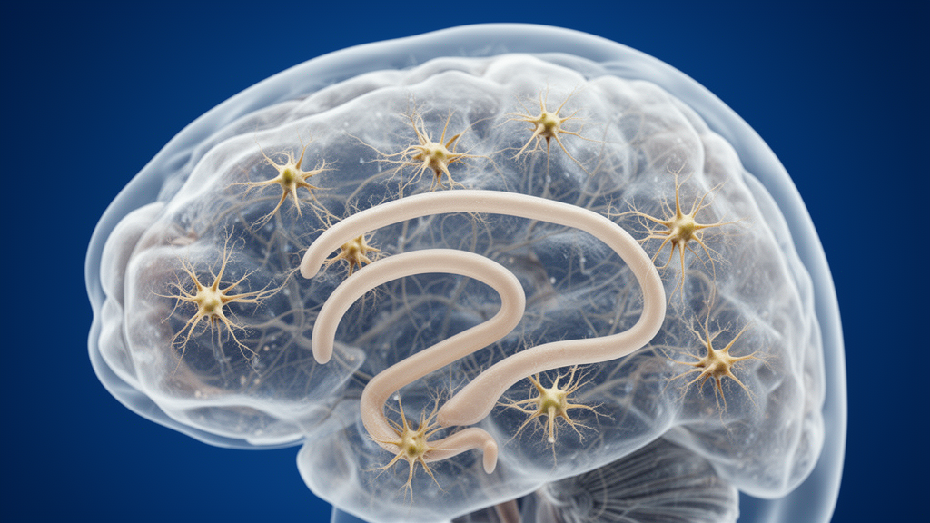
Pathological immature astroglia in the epileptic hippocampus were associated with lower fluid-intelligence scores in people with medically resistant mesial temporal lobe epilepsy, a study reports. The cells were identified by a star-like morphology and expression of doublecortin, or Dcx; the researchers used Dcx-positive cells with this morphology as a practical index of immature astroglia. Their numbers increased with age in the dentate gyrus molecular layer and were inversely associated with performance on Raven’s Progressive Matrices, a nonverbal measure of abstract reasoning and fluid intelligence. This observational study does not establish that immature astroglia cause intelligence to decline, but it identifies a cellular feature that may help researchers investigate cognitive deterioration in epilepsy.
The study, published as a brief research report in Frontiers in Cellular Neuroscience, combined preoperative neuropsychological testing with analysis of surgically removed hippocampal tissue. Mesial temporal lobe epilepsy is the most common form of adult epilepsy, and people whose seizures are not controlled with medication may undergo removal of the epileptic hippocampus. That clinical setting creates a rare opportunity to connect cognitive performance measured before surgery with cellular abnormalities in the same individuals’ brain tissue. The investigators used it to test whether an unusual astroglial population previously identified in this patient group was associated with particular cognitive deficits.
Epilepsy is associated with accelerated brain aging and progressive cognitive impairment, with cognitive decline especially pronounced in older patients. The researchers examined whether the Dcx-positive, star-shaped cells in epileptic hippocampus tracked specific domains of cognition rather than serving as a nonspecific indicator of poor overall brain health. The targets included fluid intelligence and reasoning, visuospatial reconstruction, visuospatial memory, verbal learning and other functions assessed in the preoperative evaluation. The approach was made possible because these patients often have detailed cognitive testing and, when surgery is required, provide the hippocampal tissue needed for direct cellular examination.
The cognitive cohort included 40 Spanish-speaking adults with pharmacoresistant mesial temporal lobe epilepsy. Their average age was 39.88 years, their average disease duration was 26.74 years, and they had completed an average of 7.9 years of education. The researchers retrospectively analyzed the Neuropsychological Screening Battery for Hispanics together with a grooved-pegboard fine-motor test. Raw scores were converted to education-adjusted z-scores using education-based normative data, while the pegboard measure was normalized for age and education. The 15 tests or domains covered intelligence, attention, processing speed, language, visuospatial ability, visual and verbal memory, executive functioning and fine-motor dexterity.
Among the 15 measures, the clearest age-associated declines occurred in fluid intelligence and reasoning, visuospatial reconstruction and visuospatial memory. Verbal delayed recall, visual attention, processing speed and divided attention also showed significant correlations with age in the initial analysis, but those findings did not withstand correction for multiple comparisons using the Benjamini–Hochberg false-discovery-rate procedure. Regression models then examined age alongside disease duration. The combined model was significant for fluid intelligence, although neither variable reached conventional significance independently. Age had the stronger inverse association and lower p-value, accounting for 8.3% of residual variance after adjustment for disease duration, compared with 2.7% for disease duration. Neither age nor disease duration independently predicted the two visuospatial outcomes after adjustment.
For the cellular analysis, investigators examined 29 surgically resected hippocampi. Seventeen cases came from the team’s earlier work and 12 additional cases were processed through the same staining and quantification pipeline. Dcx-positive cells with a star-like shape were counted in three regions of the dentate gyrus: the granule cell layer, the hilus and the molecular layer. Prior work by the group established that all of these cells also expressed the astroglial markers GFAP and S100β, while they lacked co-expression with markers of granule neurons, mossy cells, interneurons, microglia and PSA-NCAM-positive immature neurons. The cells were therefore treated as a practical histological index of pathological immature astroglia, and the earlier study had not detected this population in control postmortem hippocampus.
Among the 29 patients, the number of Dcx-positive immature astroglial cells increased significantly with age in the molecular layer, with p = 0.0187. Counts in the granule cell layer and hilus showed no significant age-related change. A regression model supported this regional pattern: age at surgery was a positive predictor of molecular-layer cell counts and uniquely explained 17.3% of residual variance, while disease duration was not significant and accounted for 2.7%. Age and disease duration themselves were positively correlated, but disease duration was not associated with immature-astroglia counts in any of the three regions. The result therefore points to an age-associated accumulation of the abnormal cells in one hippocampal compartment rather than a uniform increase throughout the dentate gyrus.
The direct link with cognition was tested in 19 patients for whom both Spanish-language neuropsychological data and histology were available. Greater numbers of immature astroglia in the hilus and molecular layer were associated with lower Raven’s fluid-intelligence z-scores, with p values of 0.04 and 0.02, respectively. The association in the granule cell layer approached significance at p = 0.052. These correlations did not survive correction for multiple testing, and immature-astroglia counts were not significantly associated with visuospatial reconstruction, visuospatial memory or the other tested cognitive domains. The pattern was therefore specific to abstract, nonverbal reasoning in the available data, rather than a general relationship between cell abundance and every measure of cognition.
More complex regressions produced an important qualification. When age, disease duration and immature-astroglia counts were modeled together, the overall models were significant, but none of the individual predictors remained independently significant. Age contributed the largest unique share of residual variance in fluid intelligence, from 12.6% to 25.4%, while astroglial measures contributed 8.3% to 15.6% and disease duration only 3.3% to 4.1%. Models containing age plus astroglial counts retained a negative age–intelligence relationship, whereas the astroglial counts lost significance. In models with disease duration plus astroglial counts, both were negatively associated with fluid intelligence. The authors interpret these results as evidence that age and immature astroglia share some variance in relation to cognitive performance, while also cautioning against claiming an independent effect from such a small sample.
Several limitations constrain how far the findings can be generalized. The histological sample was small, the subset with paired cognitive data included only 19 patients, and the investigators did not conduct sex-based analyses because statistical power was insufficient. Seizure frequency and other potentially relevant clinical factors could not be examined because complete seizure records were unavailable. Surgery occurred within one year of cognitive testing for 10 of the 19 paired cases, but the remaining nine underwent surgery between one and five years afterward. In addition, Dcx positivity and star-like morphology provided a practical proxy rather than a comprehensive molecular definition. The authors recommend additional astroglial markers, single-cell sequencing and spatial transcriptomics for future work to characterize these cells and the signaling pathways associated with them.
Even with those caveats, the study offers a specific new lead for studying cognitive decline in epilepsy. It links an age-related cellular change in the hippocampus with fluid intelligence, a domain that also depends heavily on frontal brain networks. Whether hippocampal immature astroglia affect reasoning through disrupted hippocampal–frontal communication, or instead accumulate as a consequence of an already deteriorating brain, remains unresolved. The work also does not show that the same process occurs in healthy aging or in other neurological disorders. Future mechanistic and larger clinical studies will be needed to determine whether the cells are a cause, a consequence or part of a broader network of changes involving seizures, neuroinflammation, oxidative stress, blood–brain barrier leakage and neuronal hyperactivity. For now, the evidence identifies immature astroglia as a candidate cellular correlate—and possible research target—rather than a proven cause of declining intelligence in people with epilepsy.
Subject of Research: Association of hippocampal immature astroglia with age-related fluid-intelligence decline in people with mesial temporal lobe epilepsy
Article Title: An age-related decline in human fluid intelligence corresponds with immature astroglia prevalence
Article References: Ammothumkandy, A., Corona, L., Atai, N. A., Abedi, A., Jimenez, N., Armacost, M., D’Orazio, L. M., Zuverza-Chavarria, V., Cayce, A., Shariq, M., McGinn, R., Lee, D., Lee, B., Heck, C., Russin, J. J., Liu, C. Y., Smith, J. A. D., & Bonaguidi, M. A. (2026). An age-related decline in human fluid intelligence corresponds with immature astroglia prevalence. Frontiers in Cellular Neuroscience, 20, Article 1926804. https://doi.org/10.3389/fncel.2026.1926804
Image Credits: AI Generated
DOI: 10.3389/fncel.2026.1926804
Keywords: astroglia, fluid intelligence, mesial temporal lobe epilepsy, cognitive decline, aging, hippocampus, doublecortin, neuropsychology, dentate gyrus, epilepsy, age-related, decline
News Source: Kendall Mcintyre. (October 20, 2026). Immature Astroglia Track Fluid-Intelligence Decline in Epilepsy Patients. Scienmag.