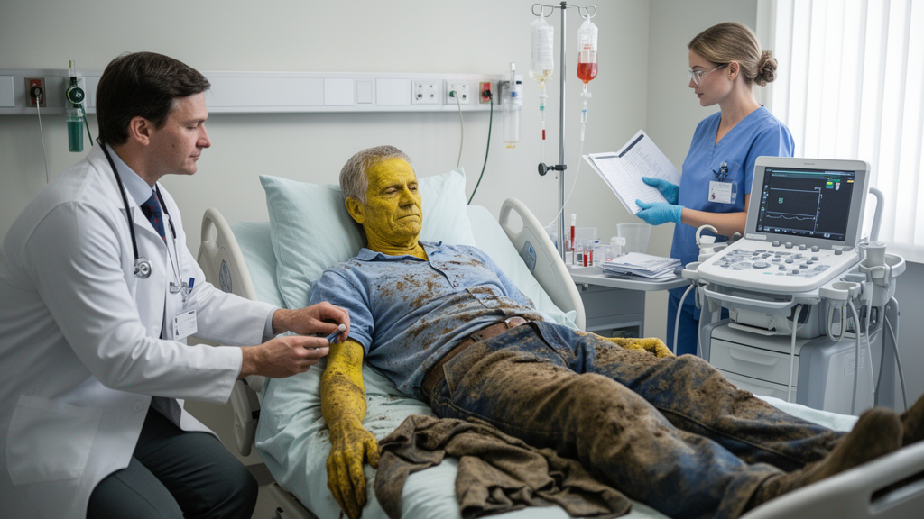
A 35-year-old farmer was admitted with fever, upper abdominal pain, jaundice and severe kidney injury in a clinical pattern that initially resembled acute cholangitis, but abdominal ultrasonography found no biliary obstruction. Testing instead identified serum anti-Leptospira IgM, while occupational exposure, calf pain, conjunctival suffusion, thrombocytopenia and multiorgan dysfunction supported severe leptospirosis presenting as Weil’s syndrome. The single-patient case report shows that leptospirosis can enter the differential diagnosis of cholestatic jaundice even when fever, abdominal pain and systemic inflammation initially point toward cholangitis.
The patient reported seven days of fever, upper abdominal pain and bilateral lower-limb pain when he arrived at the medical ward. He also had yellow-red discoloration of the sclera, dark urine, pale stools and generalized itching. His history included a 10-pack-year smoking history and reported consumption of one or two alcoholic drinks per day for five to seven years, although he had been abstinent for five months and could not recall the type of beverage, serving size or alcohol concentration. He reported no biliary colic, known gallstones or use of hepatotoxic medicines. On admission, his blood pressure was 85/50 mmHg, pulse rate 90 beats per minute, respiratory rate 28 breaths per minute, oxygen saturation 95% on room air and temperature 98.8°F. Examination showed marked icterus and conjunctival suffusion without peripheral signs of chronic liver disease. His abdomen was soft and nontender, with no right-upper-quadrant tenderness, guarding or rigidity.
Although fever, jaundice and upper abdominal pain ordinarily prompt evaluation for acute cholangitis, the patient did not meet Charcot’s triad because abdominal tenderness was absent. Initial testing showed predominantly conjugated hyperbilirubinemia: total bilirubin was 10.6 mg/dL and the direct fraction was 9.81 mg/dL. Alkaline phosphatase was elevated at 267 U/L and gamma-glutamyl transferase at 105 U/L, whereas alanine aminotransferase was mildly increased at 58.4 U/L, creating a cholestatic biochemical pattern. The total leukocyte count was 13.8 × 10^3/μL, with 92.4% neutrophils, and the platelet count was markedly reduced to 20 × 10^3/μL. The erythrocyte sedimentation rate was 95 mm/h and C-reactive protein concentration was 223 mg/L, indicating a substantial systemic inflammatory response.
Renal function was severely compromised at presentation. Blood urea was 169 mg/dL, serum creatinine was 4.97 mg/dL and the patient had oliguria, indicating acute kidney injury. Urinalysis showed two-plus protein, pus cells, mild bacteria and granular casts. Prothrombin time was 15.4 seconds, activated partial thromboplastin time 46 seconds and the international normalized ratio 1.28. Screening was negative for hepatitis B surface antigen, anti-hepatitis C virus and HIV. Dengue NS1 antigen was negative, as were malaria immunochromatographic testing and malarial parasite smear. Blood culture was negative, although additional tests for typhoid fever were not performed. A chest radiograph showed clear lung fields without consolidation or pleural effusion.
Abdominal ultrasonography identified no hepatobiliary abnormality, and the common bile duct measured 0.4 cm without evidence of obstruction. This made acute cholangitis substantially less likely because cholangitis commonly arises in association with biliary obstruction, such as a common duct stone or neoplasm. The clinicians consequently considered a wider differential that included acute cholangitis, dengue, malaria, sepsis with multiorgan failure, viral hepatitis, drug-induced liver injury and other tropical febrile illnesses. Negative viral and malaria studies weakened several alternatives, while the lack of a relevant medication history made drug-induced liver injury less likely. The combination of cholestatic jaundice, acute kidney injury, thrombocytopenia and conjunctival suffusion instead favored severe leptospirosis. His work as a farmer, repeated exposure to wet fields and prominent calf muscle pain further supported that possibility.
Leptospirosis is a globally distributed bacterial zoonosis associated particularly with populations affected by socioeconomic vulnerability. Pathogenic Leptospira species infect animal hosts such as rodents, which can shed the organisms in urine. Human infection can follow occupational or recreational contact with contaminated water or soil, with entry through abraded skin or mucous membranes. Its early presentation is often nonspecific, with fever, headache and muscle pain that can resemble dengue, influenza, malaria and other acute febrile illnesses. Approximately 10% of patients develop severe disease. The source article describes a pathogenesis involving direct tissue injury, immune-mediated mechanisms, endothelial dysfunction, disturbed microcirculation and multiorgan damage. Severe manifestations include Weil’s syndrome, classically defined by conjunctival suffusion, jaundice and acute kidney injury, as well as pulmonary, hepatic, renal, cardiac, hematological and gastrointestinal abnormalities.
A serum anti-leptospiral IgM enzyme-linked immunosorbent assay produced an index value of 2.18, above the assay’s positive threshold of 1.1. The result was considered consistent with acute leptospirosis when interpreted alongside the clinical and laboratory pattern. Confirmatory polymerase chain reaction testing for Leptospira DNA and microscopic agglutination testing were unavailable at the treating center. IgM ELISA therefore served as supportive rather than definitive confirmation in this case; the report notes that the method can cross-react and must be interpreted in an appropriate clinical context. Creatine phosphokinase activity was 39 U/L, within the laboratory’s stated reference range of 39–308 U/L.
Because the patient had severe jaundice and renal dysfunction and both leptospirosis and acute cholangitis remained under consideration, the medical team began empirical intravenous ceftriaxone twice daily with intravenous normal saline for hydration. Ceftriaxone was selected because it covers common biliary pathogens and is also an established first-line parenteral agent for severe leptospirosis. Penicillin G and doxycycline are also used for milder disease, whereas third-generation cephalosporins are preferred for severe disease with multiorgan involvement in the case report. Intravenous paracetamol was prescribed as needed for fever and pain, although the patient experienced only one episode of fever during the four-day hospital stay. He received four days of intravenous ceftriaxone in hospital and was advised to complete three additional days at a local outpatient facility, for seven days in total.
Serial laboratory measurements showed changes consistent with improvement after treatment began, although the renal trajectory was not uniformly downward on every measure. By hospital day three, the leukocyte count had fallen to 10.9 × 10^3/μL, the neutrophil percentage to 73% and total bilirubin to 6 mg/dL. Platelet count rose from 20 × 10^3/μL at admission to 22 × 10^3/μL on day one, 31 × 10^3/μL on day two and 55 × 10^3/μL on day three. Alkaline phosphatase declined from 267 U/L at admission to 250, 202 and 202 U/L on successive days. Blood urea initially rose from 169 mg/dL at admission to 196 mg/dL on day one, then fell to 162 mg/dL on day two and 85 mg/dL on day three. Serum creatinine declined across the same interval, from 4.97 to 4.5, 3.6 and 2 mg/dL. The patient remained hemodynamically stable and developed no bleeding despite severe thrombocytopenia, so platelets, fresh frozen plasma and cryoprecipitate were not given; the low platelet count was managed conservatively with close monitoring.
At follow-up one week after the antibiotic course ended, the patient reported complete resolution of fever, jaundice, abdominal pain and calf myalgia. Repeat laboratory testing showed near-normalization of bilirubin, renal parameters, leukocyte count and platelet count. Long-term follow-up was unavailable, so the report cannot establish whether renal recovery was sustained or whether chronic complications later emerged. Severe leptospirosis can produce acute kidney injury through tubulointerstitial damage, often alongside jaundice, thrombocytopenia and abnormal urine findings. Other described severe forms include pulmonary hemorrhagic syndrome, acute respiratory distress syndrome and manifestations that can meet criteria for sepsis because of intense systemic inflammation and multiorgan failure. In this patient, the combination of jaundice, acute kidney injury and hematological abnormalities corresponded to the reported pattern of Weil’s syndrome.
The central diagnostic lesson is that a cholestatic liver profile alone does not establish acute cholangitis. Although the patient had fever, upper abdominal pain, jaundice, leukocytosis and sharply elevated inflammatory markers, imaging showed no biliary obstruction, the abdominal examination did not reproduce the pain and blood cultures were negative. Severe kidney injury, thrombocytopenia, conjunctival suffusion, calf myalgia and exposure to wet agricultural fields instead formed a more coherent pattern of leptospirosis. The report supports considering the infection among the causes of suspected cholangitis when no obstruction is demonstrated, particularly in endemic settings and among people with compatible occupational exposure. As a single case without a comparator group and with limited long-term follow-up, it cannot establish comparative treatment effectiveness or population-level outcomes. Its value instead lies in documenting a severe, non-obstructive presentation and the clinical and laboratory pattern that allowed the diagnosis to be reconsidered.
Subject of Research: Severe leptospirosis presenting as Weil’s syndrome with cholestatic jaundice and multiorgan dysfunction that mimicked acute cholangitis.
Article Title: Weil's Syndrome Masquerading as Acute Cholangitis: A Case Report of a Farmer With Leptospirosis
Article References: Shaikh, A. A., Shaikh, F., Arshad, S., Devrajani, T., Ahmed, H., & Al‐Dhaheri, M. (2026). Weil's Syndrome Masquerading as Acute Cholangitis: A Case Report of a Farmer With Leptospirosis. Clinical Case Reports, 14(10), Article e73751. https://doi.org/10.1002/ccr3.73751
Image Credits: AI Generated
DOI: 10.1002/ccr3.73751
Keywords: leptospirosis, Weil’s syndrome, acute cholangitis, cholestatic jaundice, acute kidney injury, case report, thrombocytopenia, occupational exposure, bacterial zoonosis, conjugate hyperbilirubinemia, Weil's, Syndrome
News Source: Ophelia Keating. (October 20, 2026). Weil’s Syndrome Mimicked Acute Cholangitis in a Farmer With Leptospirosis. Scienmag.