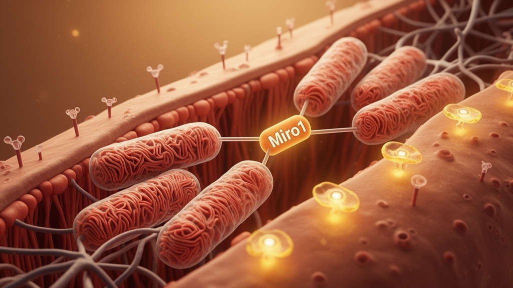
A single protein sitting on the outer membrane of mitochondria may help explain why skeletal muscle, the body’s largest glucose sink, stops responding to insulin in type 2 diabetes. In a study published in Nature Communications, researchers report that Mitochondrial Rho GTPase 1, known as Miro1, accumulates in the skeletal muscle of patients with obesity and type 2 diabetes, and that this buildup is not a passive byproduct of disease but an active participant in insulin resistance. The work, led by Elizabeth C. Heintz and John P. Kirwan at Pennington Biomedical Research Center together with Christopher L. Axelrod, combines a human cross-sectional cohort, a randomized controlled trial of exercise training, genetically engineered mice, and cell culture experiments into a single mechanistic story that links mitochondrial dynamics to glucose homeostasis.
The scale of the problem motivating the research is enormous. More than 37 million people in the United States and over 500 million people worldwide live with type 2 diabetes, and the global economic burden approaches one trillion dollars. Yet the cellular mechanisms driving the disease remain incompletely understood, which limits the development of therapies that address underlying pathophysiology rather than symptoms. Skeletal muscle is a logical place to look: it accounts for roughly 85 percent of whole-body glucose disposal at rest, and obesity, present in more than 90 percent of patients with type 2 diabetes, is the primary cause of the insulin resistance that precedes overt disease.
Miro1 is an evolutionarily conserved, atypical GTPase anchored to the outer mitochondrial membrane, with calcium-binding motifs and a cytoplasmic-facing transmembrane domain. It was first studied for its role in mitochondrial movement in neurons, where it anchors the fission machinery protein DRP1 and restricts mitochondrial transport. But earlier work hinted at broader metabolic roles: flies lacking Miro1 specifically in muscle lost the ability to climb even though their mitochondrial networks looked entirely normal. Because the researchers had previously shown that lipid-induced insulin resistance in humans involves activation of mitochondrial quality control pathways, they suspected Miro1 might behave differently in diabetic muscle than in healthy tissue.
To find out, the team analyzed muscle biopsies from three groups of patients: healthy weight individuals, people with obesity without diabetes, and people with obesity and type 2 diabetes. Each participant underwent a three-day inpatient stay with a fasting muscle biopsy and a hyperinsulinemic-euglycemic clamp, the gold-standard test for insulin sensitivity. The result was striking in its specificity. Miro1 protein accumulated in skeletal muscle mitochondria only in the patients with type 2 diabetes, not in those with obesity alone, and its abundance showed a significant negative correlation with insulin sensitivity. The pattern was replicated in mice: animals fed a high-fat, high-fructose, high-cholesterol diet, as well as monogenic diabetic models such as lethal yellow and Lepr db/db mice, all showed skeletal muscle Miro1 accumulation. Because the genetically diabetic mice were lean and ate a low-fat diet, the accumulation appeared to be driven by hyperglycemia and insulin resistance rather than by obesity or diet composition alone.
The next question was why Miro1 builds up. The researchers focused on insulin signaling at the organelle level. In the canonical pathway, insulin triggers AKT phosphorylation in the cytosol, which promotes GLUT4 transporter translocation to the cell membrane and glucose uptake. But AKT also rapidly translocates into mitochondria upon insulin stimulation, and when the team measured phosphorylated AKT at threonine 308 in isolated muscle mitochondria from patients, those with type 2 diabetes showed a near-complete absence of this mitochondrial AKT activation compared with patients who had obesity alone. In cell culture, acute insulin exposure lowered Miro1 expression, and immunofluorescence showed that AKT and Miro1 co-localization rose from low basal levels to roughly 60 percent after insulin treatment. Co-immunoprecipitation in mouse muscle confirmed a direct physical interaction between mitochondrial AKT and Miro1 that strengthened with insulin.
That interaction appears to be functionally important. When the researchers blocked AKT activity in muscle cells with the selective inhibitor MK-2206, insulin could no longer suppress Miro1, and the protein rapidly accumulated on the mitochondrial surface. In other words, insulin normally keeps Miro1 in check by delivering active AKT to mitochondria, and when that delivery fails, as it does in type 2 diabetes, Miro1 is retained and accumulates. This positions defective mitochondrial AKT translocation as a proximate cause of the Miro1 buildup observed in diabetic muscle, and it suggests that insulin resistance at the organelle level may precede and reinforce the systemic metabolic defects seen in the disease.
If defective insulin signaling causes Miro1 accumulation, then restoring insulin sensitivity should reverse it. To test this, the team conducted a randomized controlled trial, registered as NCT02977442, in which sedentary adults with obesity and type 2 diabetes were assigned to either standard medical care or a 12-week supervised aerobic exercise program of five sessions per week at moderate-to-vigorous intensity. Exercise training improved insulin sensitivity, increased GLUT4 translocation to the muscle membrane, and lowered Miro1 expression, while these measures remained unchanged in the control group. Electron microscopy revealed that exercise also relieved mitochondrial hyper-fragmentation, improving mitochondrial sphericity and elongation and significantly reducing a fragmentation index. Consistent with this remodeling, the exercisers showed reduced DRP1 phosphorylation and lower expression of Mid49 and Mid51, the adaptor proteins that recruit DRP1 to mitochondria.
The functional consequences were measurable at the level of respiratory physiology. Using high-resolution respirometry, the team found that maximal oxidative phosphorylation linked to NADH, succinate, fatty acids, complex III, and complex IV all increased after exercise training, and the creatine kinase clamp technique showed enhanced mitochondrial conductance and maximal respiratory capacity. Notably, the improvements occurred without increases in classic biogenesis markers such as PGC1α, MFN1, MFN2, OPA1, PINK1, or Parkin, suggesting that exercise remodeled the existing mitochondrial network rather than simply building more mitochondria. A reduction in LC3 expression, a marker of autophagic turnover, further hinted that the remaining mitochondria were healthier and required less quality control.
To establish causation rather than correlation, the researchers generated mice lacking Miro1 specifically in skeletal muscle, achieving roughly 70 percent reduction in protein and more than 90 percent reduction in gene expression confined to muscle tissue. When placed on a high-fat diet, these knockout mice showed lower fasting glucose, fasting insulin, and HOMA-IR than littermate controls, cleared glucose faster in response to insulin injection, and disposed of more glucose during clamps, with elevated uptake of radiolabeled 2-deoxyglucose into muscle. Insulin signaling through AKT and AS160 was enhanced, membrane-bound GLUT4 increased, and quadriceps mitochondria showed greater oxidative phosphorylation capacity and conductance. Matching experiments in C2C12 muscle cells with lentiviral Miro1 knockdown reproduced every key finding, including increased basal and insulin-stimulated glucose uptake and elevated N-linked and convergent respiratory capacity, arguing that the effects are muscle-autonomous rather than secondary to systemic adaptation.
The collective picture is that Miro1 acts as a negative regulator of skeletal muscle insulin action: insulin suppresses it through mitochondrial AKT, diabetes lets it accumulate, and its excess constrains both glucose uptake and respiratory function. Intriguingly, the authors note that the opposite pattern occurs in pancreatic beta cells, where Miro1 is depleted in diabetes and impairs insulin secretion, a tissue-specific divergence the researchers attribute to fundamentally different mitochondrial demands between hormone-secreting cells and contractile muscle. The findings position Miro1 as a novel link between mitochondrial quality control and insulin resistance, and they suggest that pharmacologically targeting Miro1, or mimicking the exercise-induced reduction in its abundance, could offer a new therapeutic avenue for restoring insulin sensitivity in type 2 diabetes. The authors caution that prospective human studies are still needed to track Miro1 as patients transition from obesity to overt diabetes, and that direct modulation of Miro1 may also affect muscle contractility, an important consideration for any future intervention.
Subject of Research: The role of the outer mitochondrial membrane protein Miro1 in skeletal muscle insulin resistance and glucose homeostasis in type 2 diabetes
Article Title: Miro1 mediates skeletal muscle insulin resistance in type 2 diabetes
Article References: Heintz, E. C., Dantas, W. S., Taylor, A. L., Zunica, E. R. M., Belmont, K. P., Slone, J. M., Mey, J. T., Beyl, R., Vandanmagsar, B., Parry, H. A., Ajayi, P., Glancy, B., Axelrod, C. L., & Kirwan, J. P. (2026). Miro1 mediates skeletal muscle insulin resistance in type 2 diabetes. Nature Communications, 17(1), Article 10702. https://doi.org/10.1038/s41467-026-77278-6
Image Credits: AI Generated
DOI: 10.1038/s41467-026-77278-6
Keywords: Miro1, type 2 diabetes, insulin resistance, skeletal muscle, mitochondrial dynamics, AKT signaling, exercise training, GLUT4, mitochondrial fission, DRP1, glucose homeostasis, oxidative phosphorylation
News Source: Ophelia Keating. (October 19, 2026). Mitochondrial Protein Miro1 Emerges as a Driver of Muscle Insulin Resistance in Type 2 Diabetes. Scienmag.