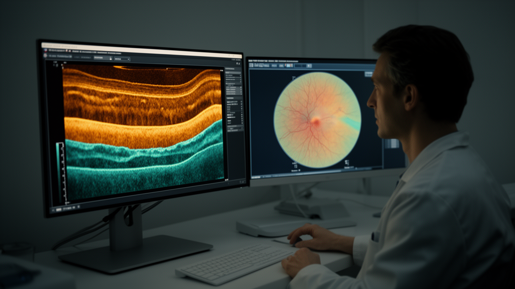
Optical coherence tomography has become one of the most widely used imaging tools in ophthalmology, offering non-invasive cross-sectional views of the retina at micrometer resolution. Yet the clinical value of these scans depends on how reliably they can be interpreted, and the growing volume of OCT examinations worldwide has intensified interest in automated diagnosis. A new study published in Complex & Intelligent Systems presents an explainable, topology-aware frequency-semantic transformer that pushes multi-class retinal OCT classification to a reported accuracy of 97.86 percent, outperforming a range of established convolutional and transformer-based baselines.
The research, conducted by Jeena Joseph of the Department of Computer Applications at Marian College Kuttikkanam Autonomous in Kerala, India, addresses a problem that has long frustrated machine learning approaches to retinal imaging: the subtle differences within disease classes, the visual similarities between different retinal conditions, and the complex anatomical deformations that disease itself produces. These factors make accurate multi-class OCT diagnosis genuinely difficult, because a model must simultaneously distinguish fine-grained texture changes and preserve awareness of the retina’s layered global structure.
The framework described in the paper combines three complementary strands of representation learning. Spatial semantic learning captures the local appearance of retinal features in the image domain. Frequency-aware representation extraction operates in the frequency domain, where periodic structures such as the alternating bright and dark bands of retinal layers produce characteristic spectral signatures that can reveal disruptions invisible to purely spatial analysis. Topology-preserving structural modeling then encodes the continuity of retinal anatomy, allowing the model to register local violations, such as fluid pockets or layer breaks, against the expectation of global anatomical continuity.
At the heart of the architecture is a cross-scale transformer interaction module designed to improve semantic coordination between fine-grained retinal textures and large-scale structural representations. Transformers process information through attention mechanisms that weigh the relevance of every part of an input to every other part, and by operating across scales the module lets high-resolution detail inform coarse anatomical reasoning and vice versa. This cross-talk is what enables the network to connect a small intraretinal lesion to its position within the broader layered architecture of the retina, a relationship that matters clinically because the same disease can manifest differently depending on which layer is affected.
The second distinguishing component is an explainability-guided attention module. Rather than treating interpretability as an afterthought applied once a model has been trained, the study incorporates it directly into the learning process to enhance lesion localization and the interpretability of the model’s decisions. In medical imaging, this matters enormously: a classifier that reaches the right answer for the wrong reasons may perform well on benchmark metrics while failing catastrophically on unseen data or, worse, on patients whose presentations differ from the training population. Attention that can be inspected and validated against known lesion locations gives clinicians a basis for trusting, or questioning, the model’s output.
Evaluation was carried out on the Retinal OCT-C8 dataset, a benchmark comprising 24,000 OCT images spanning eight retinal disease categories. The proposed framework achieved a classification accuracy of 97.86 percent, precision of 97.53 percent, recall of 97.21 percent, an F1-score of 97.36 percent, and an area under the receiver operating characteristic curve of 99.02 percent. These figures place the model ahead of state-of-the-art CNN- and transformer-based competitors reported in the study, with the margin quantified precisely: a 1.48 percentage-point improvement in accuracy over the strongest CNN-transformer hybrid baseline and a 5.02 percentage-point improvement over ResNet50, a widely used convolutional architecture that has served as a standard reference point in medical image classification for years.
The comparison to ResNet50 is instructive because it illustrates a broader shift in the field. Convolutional networks excel at local pattern recognition but process information through fixed receptive fields, which can make it harder to capture long-range dependencies across an image. Transformers, by contrast, attend globally, but vanilla vision transformers can struggle with the dense, repetitive texture patterns characteristic of OCT scans. Hybrid approaches attempt to combine the strengths of both, and the fact that the new framework still improves on the strongest hybrid baseline by nearly one and a half percentage points suggests that the gains come not merely from architecture choice but from the explicit encoding of frequency and topological information that neither paradigm captures on its own.
Beyond raw accuracy, the author reports that the framework enhances robustness, cross-domain generalization, and explainability. Cross-domain generalization is a persistent challenge in medical AI, because models trained on scans from one imaging device or patient population often degrade when applied to another. Robustness to such shifts is what separates a laboratory demonstration from a deployable clinical tool, and the study’s emphasis on structural and frequency-based representations, which are less tied to device-specific appearance than raw pixel statistics, offers a plausible mechanism for that resilience.
The clinical stakes are considerable. Retinal diseases, including various forms of macular degeneration, diabetic macular edema, and other conditions represented in multi-class OCT benchmarks, are among the leading causes of vision loss worldwide, and early detection can determine whether abnormalities that threaten sight are caught in time. An automated system that reliably distinguishes among eight disease categories while flagging the specific regions that drove its decision could support screening programs, triage urgent cases, and reduce the interpretive burden on ophthalmologists facing ever-growing imaging queues.
The study used a publicly available dataset with no direct human or animal involvement, and the author reports no funding and no conflicts of interest. The work was received in July 2026, accepted in October 2026, and published on 18 October 2026 under open access terms. As deep learning continues its advance into diagnostic medicine, this research offers a concrete example of how combining domain knowledge about retinal anatomy, signal-processing insights from the frequency domain, and built-in explainability can yield models that are not only more accurate but more transparent, a combination that will be essential if artificial intelligence is to earn a lasting role in the clinic.
Subject of Research: Explainable topology-aware frequency-semantic transformer for multi-class retinal OCT diagnosis
Article Title: Explainable topology-aware frequency-semantic transformer for robust multi-class retinal OCT diagnosis
Article References: Joseph, J. (2026). Explainable topology-aware frequency-semantic transformer for robust multi-class retinal OCT diagnosis. Complex & Intelligent Systems. https://doi.org/10.1007/s40747-026-02571-2
Image Credits: AI Generated
DOI: 10.1007/s40747-026-02571-2
Keywords: retinal OCT, deep learning, vision transformer, explainable AI, topology-aware learning, frequency-semantic representation, retinal disease classification, medical imaging, attention mechanism, cross-domain generalization, Complex & Intelligent Systems, lesion localization
News Source: Ophelia Keating. (October 18, 2026). AI Reads Retinal Scans With Frequency and Topology Awareness for Sharper OCT Diagnosis. Scienmag.