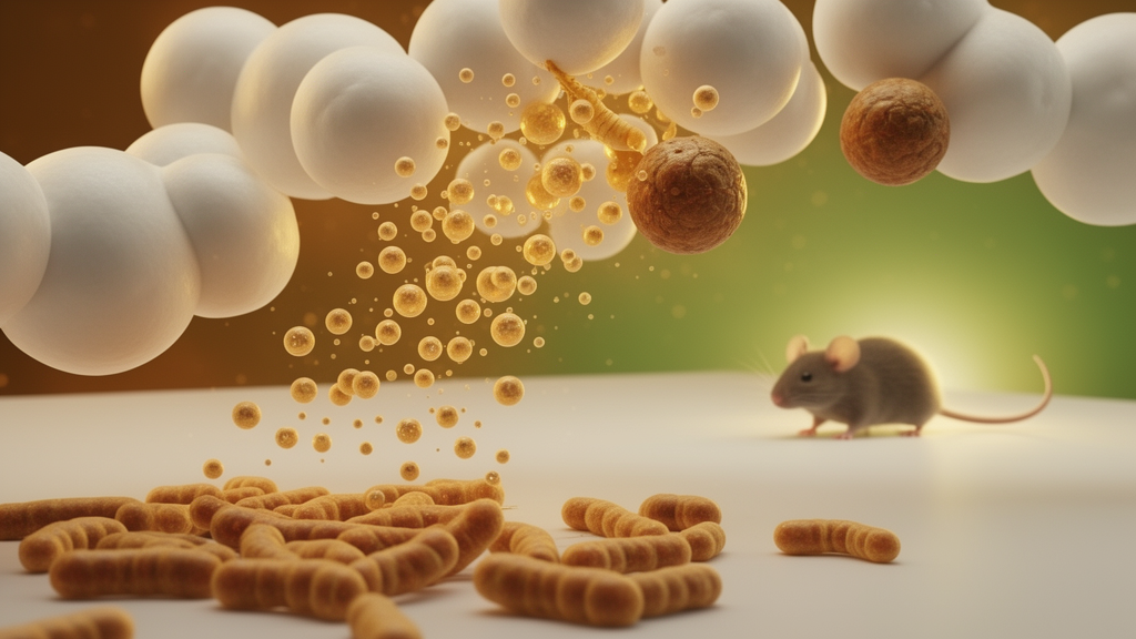
Tiny vesicles released by ginseng roots may be doing more than carrying plant chemistry across the gut lining. In a new mouse study, researchers at Sun Moon University in Korea report that ginseng-derived extracellular vesicles, or GDE, given orally to mice on a high-fat diet produced measurable changes in fat tissue structure, restored responsiveness to a key satiety hormone, and shifted the composition of the gut microbiome. The findings, published in Scientific Reports, add to a growing body of work suggesting that plant-derived nanovesicles are not inert dietary debris but biologically active particles capable of influencing host metabolism.
Extracellular vesicles are lipid-bilayer-bound nanoparticles that cells, including plant cells, naturally release into their surroundings. In recent years, scientists have isolated similar vesicles from fruits, vegetables, and medicinal herbs and found that they can survive digestion, cross intestinal barriers, and deliver cargo such as proteins, lipids, and small RNAs to recipient cells. Panax ginseng, a root long used in traditional East Asian medicine and increasingly studied for its ginsenoside content, has emerged as a source of such vesicles. But while in vitro studies have hinted at anti-inflammatory and metabolic effects, the team behind the new study, led by corresponding author Sunhye Lee, wanted to know what happens when these vesicles are actually fed to living animals under controlled dietary conditions.
The experimental design was straightforward but rigorous. Male C57BL/6 mice, a standard laboratory strain widely used in obesity research, were divided into three groups. One group received a low-fat diet, serving as a healthy baseline. A second group received a high-fat diet designed to induce obesity. A third group received the same high-fat diet but was also given GDE orally three times per week, at a dose of 3 billion particles per administration, for nine weeks. This dosing schedule allowed the researchers to track cumulative effects of the vesicles over a substantial portion of the animals’ adult lives without relying on injections or invasive delivery methods.
After nine weeks, the differences between the high-fat diet group and the high-fat diet plus GDE group were striking in several respects. The epididymal fat pads, a common site of visceral fat accumulation in male mice, showed reduced relative weight in the GDE-treated animals compared to untreated high-fat diet controls. Histological examination of adipose tissue revealed that individual adipocytes, the fat-storing cells that balloon in size during obesity, were smaller in diameter in the GDE group. This matters because adipocyte hypertrophy, the pathological enlargement of fat cells, is closely linked to insulin resistance, chronic inflammation, and dysfunctional fat tissue remodeling. Notably, the researchers found that leptin mRNA expression in epididymal fat, which tends to rise as fat cells enlarge, was normalized in the GDE-treated mice. Importantly, energy intake was comparable between the high-fat diet group and the GDE-supplemented group, meaning the observed differences in fat tissue were not simply a byproduct of the treated animals eating less overall.
One of the most intriguing findings involved cholecystokinin, or CCK, a peptide hormone released by cells in the small intestine in response to food intake. CCK binds to receptors in the gut and brain, triggering sensations of fullness and slowing gastric emptying. In obesity, this satiety signaling pathway can become blunted, meaning the same amount of CCK produces a weaker feeling of satiety, potentially contributing to overeating. To test whether GDE affected this pathway, the researchers administered exogenous CCK to the mice and measured how much food they subsequently consumed. Mice on the high-fat diet alone showed a significantly reduced anorexigenic response, meaning CCK did not suppress their food intake as effectively as it did in the low-fat diet controls. However, mice that had received GDE alongside the high-fat diet showed a restored, significant suppression of food intake in response to exogenous CCK. This suggests that the vesicles may have helped preserve or restore the sensitivity of the satiety signaling pathway even in the context of an obesogenic diet.
The researchers also turned their attention to the gut microbiome, the complex community of bacteria residing in the intestines that has been increasingly implicated in obesity, inflammation, and metabolic health. Using sequencing-based analysis, they found that GDE administration altered the composition of the gut microbial community in high-fat diet-fed mice. Specifically, the relative abundance of three bacterial genera, Blautia, Colidextribacter, and Alistipes, decreased in the GDE-treated group, while two other genera, Anaerotruncus and Parabacteroides, increased. These shifts are notable because several of these genera have been previously associated with metabolic phenotypes in other studies, though the precise functional consequences of these particular compositional changes remain an open question that the authors acknowledge requires further investigation.
Inflammation in the intestinal wall is another factor that has been linked to diet-induced obesity and metabolic dysfunction. The researchers measured the expression of tumor necrosis factor-alpha, or TNF-alpha, a pro-inflammatory cytokine, in the ileum, the final section of the small intestine. They found that TNF-alpha expression was elevated in mice fed the high-fat diet alone but attenuated in mice that also received GDE. This suggests that the vesicles may have had an anti-inflammatory effect locally in the gut. However, when the researchers measured plasma levels of lipopolysaccharide-binding protein, a circulating marker of bacterial endotoxin exposure and systemic inflammation, they found no significant differences between groups. This indicates that the anti-inflammatory effects of GDE may have been localized to the intestinal tissue rather than producing broad systemic changes detectable in the bloodstream.
The study’s findings collectively point to a multi-layered mechanism by which ginseng-derived extracellular vesicles might influence diet-induced obesity. Rather than acting through a single pathway, the vesicles appear to simultaneously affect adipose tissue morphology, satiety hormone responsiveness, gut microbial composition, and local intestinal inflammation. This kind of pleiotropic effect is consistent with the complex nature of obesity as a disease that involves dysregulation across multiple organ systems and signaling networks. It also aligns with the broader hypothesis in the field that dietary bioactive vesicles can act as signaling agents that modulate host physiology through the gut environment.
The authors are careful to note the limitations of their work. Only male mice were included in the study, which means the findings cannot be directly extrapolated to females without further research. Additionally, while the study demonstrates associations between GDE administration and changes in adipocyte size, CCK responsiveness, and microbiome composition, the precise molecular mechanisms linking these observations remain to be fully characterized. The researchers state that further investigation is needed to understand how the vesicles interact with intestinal cells, whether their effects are mediated by specific cargo molecules, and whether the microbiome changes are a cause or consequence of the metabolic improvements observed.
Despite these caveats, the study represents a meaningful step forward in the emerging field of plant-derived extracellular vesicle research. By demonstrating in vivo effects on multiple obesity-related parameters using an orally administered, naturally sourced nanoparticle preparation, the work provides a foundation for future studies exploring whether similar vesicles from other plants, or purified fractions of GDE itself, could be developed as dietary interventions for metabolic health. The research was supported by a 2024 grant from The Korean Society of Ginseng, and the findings are published as open access, allowing other researchers to build upon the results. As interest in the gut-adipose-brain axis continues to grow, studies like this one highlight the potential of plant nanovesicles as tools for understanding and potentially modulating the complex biology of obesity.
Subject of Research: Effects of orally administered ginseng-derived extracellular vesicles on adipocyte morphology, satiety signaling, gut microbiota, and intestinal inflammation in diet-induced obese mice
Article Title: Ginseng derived extracellular vesicles attenuate adipocyte hypertrophy and modulate gut microbiota and cholecystokinin responsiveness in high fat diet fed mice
Article References: Jeong, E., Chae, S., Chai, H., Kim, J. W., & Lee, S. (2026). Ginseng derived extracellular vesicles attenuate adipocyte hypertrophy and modulate gut microbiota and cholecystokinin responsiveness in high fat diet fed mice. Scientific Reports. https://doi.org/10.1038/s41598-026-74734-7
Image Credits: AI Generated
DOI: 10.1038/s41598-026-74734-7
Keywords: ginseng, extracellular vesicles, adipocyte hypertrophy, cholecystokinin, gut microbiota, diet-induced obesity, Panax ginseng, leptin, intestinal inflammation, C57BL/6 mice, satiety signaling, plant-derived nanoparticles
News Source: Daisy Hatcher. (October 18, 2026). Ginseng Nanovesicles Reshape Fat Cells, Gut Microbes, and Satiety Signals in Obese Mice. Scienmag.