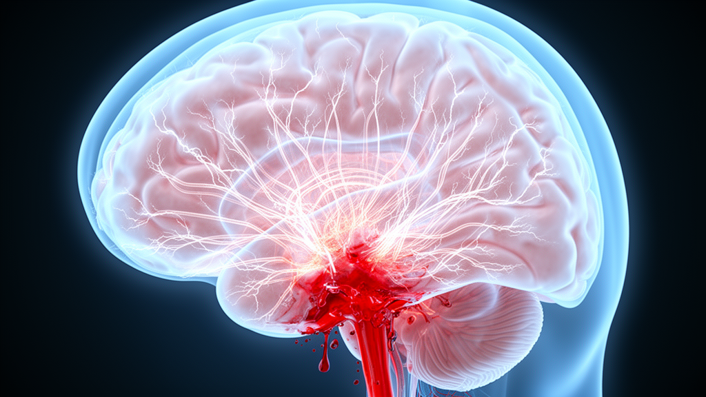
When a brain aneurysm ruptures, the bleeding itself is only the first blow. Within days, many patients develop a second, often more insidious threat: global cerebral edema, a diffuse swelling of the brain that can crush delicate tissue against the skull. Now, a team of neurosurgeons and neurointensivists has proposed a provocative new explanation for why this swelling happens, pointing the finger not at the blood alone, but at the body’s own autonomic nervous system, the vast network that governs heart rate, blood pressure, and blood-vessel tone without our awareness.
The hypothesis, published as a viewpoint in the journal Neurocritical Care by Siddharth Srinivasan, Mithun Sattur, Kautilya Patel, and Daryl R. Gress, introduces the term neurogenic cerebral edema. The idea is deliberately modeled on a better-known phenomenon: neurogenic pulmonary edema, a sudden flooding of the lungs that strikes a subset of patients after the same kind of brain hemorrhage. If autonomic chaos can wreck the delicate barrier in the lungs, the authors argue, perhaps it can also amplify the breakdown of the blood–brain barrier, allowing protein-rich fluid to seep into brain tissue and drive the vasogenic component of cerebral edema.
The clinical stakes are considerable. Global cerebral edema occurs in up to 67 percent of cases of aneurysmal subarachnoid hemorrhage, typically emerging within the first three days after the bleed, and it stands as an independent predictor of both long-term disability and death at three months. The moment of rupture brings an abrupt spike in intracranial pressure that can transiently halt the brain’s circulation altogether, setting off a cascade of energy failure, disrupted calcium handling, and mitochondrial dysfunction inside cells. Microdialysis studies in affected patients reveal metabolic distress, with abnormal lactate–pyruvate ratios and depleted glucose. Yet these well-documented mechanisms have never fully explained the diffuse, malignant swelling seen in the sickest patients.
The authors’ proposal does not discard the established pathways. Instead, they suggest that edema after hemorrhage is a mixed phenomenon, combining cytotoxic swelling inside cells, ionic edema in which ions and water cross a relatively intact barrier, and vasogenic edema that follows true blood–brain barrier injury. What the autonomic nervous system adds, in their framing, is an amplifier. Autonomic disequilibrium, they propose, may indirectly intensify known barrier-injury pathways, including the upregulation of the SUR1-TRPM4 channel in endothelial cells, neurons, and astrocytes, and the disorganization of tight junctions that normally seal the barrier shut.
The anatomical substrate for such an effect is real. The central autonomic network, first described by Benarroch in 1993, spans the medial prefrontal cortex, cingulate cortex, insula, amygdala, hypothalamus, thalamus, and key brainstem regions. Postmortem analyses and diffusion tensor imaging have shown that subarachnoid hemorrhage can directly injure the hypothalamus, igniting the sympathetic–adrenomedullary axis and unleashing a surge of catecholamines. Meanwhile, the cerebral circulation is extraordinarily rich in parasympathetic fibers, carrying ten to forty times more parasympathetic innervation than vessels outside the skull. Unusually, these intracranial parasympathetics rarely use acetylcholine as their main transmitter; instead they release potent vasodilators such as vasoactive intestinal peptide, pituitary adenylate cyclase-activating peptide, and nitric oxide.
Experimental work lends the concept biological plausibility, though the authors are careful about its limits. Stimulating the sphenopalatine ganglion, a cranial parasympathetic relay, improves cortical perfusion in rodents and can ease vasospasm in primates after experimental hemorrhage, but in those same primate experiments it transiently increased blood–brain barrier permeability, and the investigators warned that prolonged stimulation might hold the barrier open and worsen vasogenic edema. On the sympathetic side, blocking the superior cervical ganglion in mice improved cerebral microcirculation and reduced edema after hemorrhage. Heart-rate variability studies in patients reveal a striking picture of sympathovagal imbalance, with biochemical evidence of catecholamine excess coexisting with a shift toward parasympathetic predominance, a disequilibrium rather than a simple one-sided overdrive.
The analogy with the lungs is the conceptual heart of the paper. In neurogenic pulmonary edema, which affects roughly 2 to 31 percent of subarachnoid hemorrhage patients, two mechanisms are classically described: a hemodynamic pathway in which sympathetic activation raises intravascular pressure and forces fluid into the pulmonary interstitium, and a permeability pathway in which neurally driven catecholamines and cytokines loosen the endothelial barrier. Analysis of edema fluid from patients supports both hydrostatic and permeability contributions. The cerebral circulation shares dense autonomic innervation and tight-junction-dependent barrier integrity with the pulmonary vasculature, and the authors envision a parallel process in the brain: autonomic forces converging on an injured barrier, amplifying the leakage that produces vasogenic swelling. The heart, too, belongs in this picture, since neurogenic stunned myocardium after hemorrhage can reduce cardiac output and destabilize blood pressure, secondarily stressing cerebral perfusion. The brain, lungs, and heart, they argue, are organ-specific targets of a shared central stress response.
Supporting imaging evidence comes from magnetic resonance studies performed within the first 72 hours after hemorrhage. While low apparent diffusion coefficient values signal restricted diffusion from cellular injury, several studies have found the opposite: globally elevated ADC values, even in brain regions that look normal on conventional sequences, consistent with diffuse extracellular water accumulation and blood–brain barrier dysfunction. The authors illustrate the phenotype with a 26-year-old woman who suffered a poor-grade hemorrhage from a ruptured anterior communicating artery aneurysm and developed malignant cerebral edema within 48 hours. Her diffusion MRI showed diffusely elevated ADC values, including 1338 and 1297 × 10⁻⁶ mm²/s in bilateral temporal cortex and 1441 × 10⁻⁶ mm²/s in a left frontal lesion, against a contralateral white-matter reference of 928 × 10⁻⁶ mm²/s, an ADC ratio of 1.55 supportive of vasogenic edema. She required pentobarbital coma and ultimately decompressive craniectomy to control intracranial hypertension. Crucially, the authors concede that no autonomic measurements or direct barrier-permeability assessments were obtained in her case, so it demonstrates a vasogenic component without proving a neurogenic cause.
That honesty defines the paper’s tone. The authors explicitly state that neurogenic cerebral edema is a testable mechanistic hypothesis, not a diagnostic entity, and that it does not replace ischemic injury as an explanation. They distinguish it from paroxysmal sympathetic hyperactivity, a consensus-defined clinical syndrome, and they caution that experimental ganglion stimulation does not establish that spontaneous dysautonomia after hemorrhage reproduces the intensity or distribution of electrical stimulation. Their proposed validation strategy is concrete: baseline computed tomography with the Subarachnoid Hemorrhage Early Brain Edema Score at presentation, continuous or repeated autonomic measures such as heart-rate variability, catecholamines, and blood-pressure variability in prespecified early time windows, and serial imaging with direct blood–brain barrier permeability assessment over the following 6 to 24 hours, tracked through 72 hours. Machine-learning approaches could then test whether autonomic burden predicts malignant edema progression while accounting for intensive care confounders such as sedation, ventilation, and vasoactive drugs.
If validated, the framework could open therapeutic avenues aimed at preserving both barrier integrity and autonomic stability, from neuromodulation, already being explored in a randomized trial of transcutaneous auricular vagus nerve stimulation that increased parasympathetic outflow in hemorrhage patients, to pharmacological strategies targeting SUR1-TRPM4, whose inhibition with glibenclamide has reduced vasogenic edema and inflammation in experimental models. For now, the hypothesis remains exactly that: a coherent, testable story that reframes the brain’s most feared swelling as, in part, a disease of the nerves that regulate it. Given that cerebral edema remains a major determinant of outcome after aneurysmal hemorrhage, and that its mechanisms remain incompletely characterized, the proposal offers researchers something they have lacked, a specific, time-ordered set of measurements that could finally connect the storm in the autonomic nervous system to the flood in the brain.
Subject of Research: Proposed role of autonomic dysfunction in vasogenic cerebral edema after aneurysmal subarachnoid hemorrhage
Article Title: Neurogenic Cerebral Edema: Proposed Mechanism for Vasogenic Cerebral Edema
Article References: Srinivasan, S., Sattur, M., Patel, K., & Gress, D. R. (2026). Neurogenic Cerebral Edema: Proposed Mechanism for Vasogenic Cerebral Edema. Neurocritical Care. https://doi.org/10.1007/s12028-026-02684-z
Image Credits: AI Generated
DOI: 10.1007/s12028-026-02684-z
Keywords: aneurysmal subarachnoid hemorrhage, global cerebral edema, neurogenic cerebral edema, blood–brain barrier, autonomic nervous system, vasogenic edema, sympathetic activation, parasympathetic dysfunction, neurogenic pulmonary edema, SUR1-TRPM4, apparent diffusion coefficient, neurocritical care
News Source: Cassandra Pierce. (October 15, 2026). A Runaway Nervous System May Drive Deadly Brain Swelling After Hemorrhagic Stroke. Scienmag.