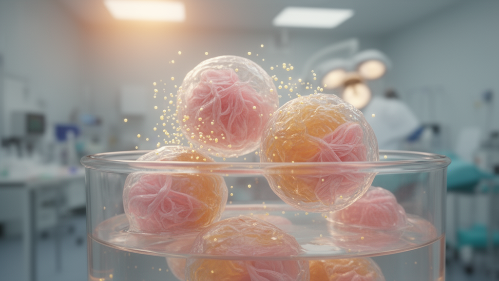
One of the most stubborn complications in chest surgery may finally have a biological answer. When surgeons remove part of a lung, the thin membrane that wraps the organ, the visceral pleura, often fails to heal completely. Air then escapes from the lung surface into the chest cavity, a condition known as persistent pulmonary air leakage that strikes between 10 and 26 percent of patients after thoracic surgery. The consequence is prolonged hospitalization, elevated infection risk and mounting healthcare costs. Now, a research team reporting in Experimental & Molecular Medicine has grown the first organoids of the pleura and demonstrated that transplanting them into damaged pleural tissue can rebuild the membrane’s cellular architecture, restore its barrier function and quiet the inflammatory storm that follows injury.
Current remedies for air leaks are largely mechanical. Synthetic polymer patches, biological grafts such as bovine pericardium and chemical pleurodesis with talc or bleomycin all aim to physically seal the defect or provoke inflammatory adhesions that glue the lung to the chest wall. These measures offer temporary relief but frequently aggravate the problem, triggering foreign body reactions, chronic inflammation and fibrosis that can produce recurrent leaks or pleural adhesions. What has been missing is a therapy that not only plugs the hole but regenerates the pleura’s native structure: a superficial mesothelial monolayer, submesothelial connective tissue and elastic fiber networks that together maintain pulmonary integrity through every breath.
Organoid technology offered the researchers a way forward. Organoids are self-organizing three-dimensional tissue mimics grown from stem cells that replicate the structural and functional complexity of their parent tissue. The approach has already delivered pancreatic islet organoids that restore glucose homeostasis in diabetic models, intestinal organoids that enhance epithelial regeneration after ischemia-reperfusion injury, and liver organoids that mitigate fibrosis by repolarizing macrophages toward a proregenerative phenotype. Yet no one had applied the technology to the pleura, despite its unique mesothelial-stromal interface and its vulnerability to mechanical stress during respiratory cycles.
The team, working at Guangdong Medical University, began with mouse lungs. To avoid contamination with deeper parenchymal cells, they isolated intact lungs without cutting, tied off the trachea and suspended the tissue in collagenase solution, carefully controlling digestion between 30 and 60 minutes. Cells recovered from the pleural surface were embedded in Matrigel and cultured in a medium tuned for mesothelial growth, containing R-spondin, EGF, FGF-10 and Noggin alongside pleural maturation additives such as the TGF-beta receptor inhibitor A83-01, ascorbic acid, retinoic acid and dexamethasone, while reducing the epithelial-supporting factors that drive alveolar and airway differentiation. Within a week, the pleural cells formed organoids with thick walls and a central lumen, clearly distinct from the honeycomb-like alveolar clusters produced by matched lung organoids grown from the same animals.
Molecular profiling confirmed the distinction. Pleura organoids expressed markedly less EPCAM, the pan-epithelial marker, and fewer alveolar markers such as AQP5 and SFTPC or airway markers such as CC10 and p63. Instead, they upregulated mesothelin (MSLN) and calretinin, canonical mesothelial markers, along with vimentin, a stromal and fibroblast marker. Enzyme-linked immunosorbent assays showed that the pleura organoids secreted higher levels of phosphatidylcholine and hyaluronic acid, lubricating substances typically produced by mesothelial cells to keep the pleural cavity gliding smoothly. The organoids retained strong Ki67-positive proliferative activity and could be passaged for more than 20 consecutive passages, a scalability that matters for any future clinical application. The protocol adapted readily to human tissue: pleura-covered areas of surgically resected healthy lung, digested with the pleural surface just submerged in collagenase, yielded human organoids with the same thick-walled morphology, mesothelial marker expression and secretory function, sustained by additions such as nicotinamide, prostaglandin E2 and the GSK3 inhibitor CHIR99021.
To test whether the organoids could form pleura-like structures in a living host, the researchers transplanted human pleura organoid cells beneath the kidney capsule of immunodeficient NSG mice. Three weeks later, a well-organized multilayered structure had formed at the graft site, closely resembling the superficial pleural region of human lung tissue. Verhoeff staining revealed organized collagen and elastic fibers, key components of the visceral pleura, and staining for human nuclear antigen confirmed the structures were built by the transplanted cells rather than by mouse tissue. Crucially, the outermost layer consisted of a continuous sheet of mesothelial cells, validating the structural fidelity of the graft.
Functional testing followed. In Transwell wound-healing assays, adding pleura organoid cells to the upper compartment accelerated the closure of scratches in mesothelial monolayers, even when the inflammatory cytokine TNF-alpha was added to mimic the environment of a fresh wound. The researchers then turned to precision-cut lung slices, 200-micrometer sections of intact lung that preserve the visceral pleura and cellular diversity. Notches cut into the pleural surface simulated pleural rupture, and fluorescently labeled human organoid cells were applied dropwise to the injury. Confocal imaging showed the transplanted cells integrating into the damaged area and, within seven days, forming a continuous, dense, multilayered pleura-like structure on the lung surface composed primarily of mesothelin-positive cells.
The decisive experiment came in living animals. The team punctured the visceral pleura of NSG mice through the chest wall to simulate surgical rupture, then delivered human pleura organoid cells into the pleural cavity. Over 35 days, the transplanted mice gained weight steadily while control mice, which received the carrier medium without cells, lost weight before slowly recovering. Histology showed organoid integration into the lung surface as early as day seven, complete filling of the wound, maturation into a dense multilayered structure by day 21 and progressive remodeling into a looser two-to-three-layered organization by day 35, approaching the single-layer architecture of the native pleura. Immunostaining for mesothelin revealed a continuous mesothelial layer on the lung surface by day seven that remained intact and integrated with the host mesothelium through day 35, and co-localization with human nuclear antigen proved the new lining derived from the graft. Barrier function, quantified by injecting fluid into the trachea and measuring the blast volume, the critical threshold at which fluid leaks past the pleura, was significantly higher in transplanted mice at every time point. Inflammation also subsided: TNF-alpha and IL-6 in alveolar lavage fluid, elevated in injured controls even at day 35, approached normal levels in transplanted animals, and expression of alpha-SMA, a fibrosis marker, was significantly attenuated. Histopathology of the heart, liver, spleen and kidney showed no lesions, indicating the therapy was well tolerated.
Mechanistically, the researchers compared organoids that had engrafted and participated in repair with those that had not. Transcriptome sequencing revealed that CD133, also known as PROM1, a stem and progenitor marker never before investigated in mesothelial contexts, was highly expressed in the transplanted organoids. Flow-sorted CD133-positive cells formed larger and more numerous organoids than CD133-negative cells, and the organoids they generated contained mesothelial, epithelial and stromal lineages, demonstrating multilineage potential, whereas CD133-negative cells differentiated only into epithelial cells. RNA sequencing identified 1,457 differentially expressed genes between the populations, with CD133-positive cells upregulating stemness-associated genes and enriching pathways tied to cell division and distinct metabolic programs. A simplified lineage-tracing experiment tracked mesothelin expression over seven days: CD133-positive organoids showed minimal mesothelin on day one, detectable levels by day three and widespread expression by days five and seven, while CD133-negative cells almost completely failed to become mesothelial cells under identical conditions. Single-cell RNA sequencing of more than 116,000 human lung cells independently confirmed a distinct CD133-positive stem cell subpopulation, positioned at a terminal point of a pseudotime differentiation trajectory, with CD133 expression progressively increasing along the trajectory, and immunostaining localized CD133-positive cells along and adjacent to the pleura in both human and mouse lung under homeostatic conditions.
When CD133-positive and CD133-negative organoids were transplanted into mice with pleural rupture, the difference was stark. CD133-positive grafts nearly maintained body weight, formed larger grafts that continuously covered the pleural wound, markedly increased blast volumes, remodeled collagen and elastic fibers and significantly reduced TNF-alpha and IL-6, while CD133-negative grafts showed only limited repair. In the regenerated tissue, CD133-positive cells co-expressed mesothelin in the surface layer, suggesting direct contribution to mesothelial formation, although the authors note that definitive in vivo lineage tracing is still needed to confirm direct differentiation. The work establishes the first pleura organoid models, identifies CD133-positive cells as a previously unrecognized regenerative population and offers a proof of concept for a progenitor-driven therapy that could one day be personalized: pleural biopsies from patients could generate autologous organoids, mitigating immune rejection while restoring pulmonary homeostasis after surgery.
Subject of Research: Regenerative Medicine
Article Title: Pleura organoid transplantation facilitates visceral pleural repair through CD133+ progenitor cell-mediated regeneration
Article References: Deng, B., Huang, P., Wang, Q., He, Y., Gong, L., Chen, C., Wu, Y., Lu, Z., & Liang, Z. (2026). Pleura organoid transplantation facilitates visceral pleural repair through CD133+ progenitor cell-mediated regeneration. Experimental & Molecular Medicine, 58(9), 2990-3006. https://doi.org/10.1038/s12276-026-01825-w
Image Credits: AI Generated
DOI: 10.1038/s12276-026-01825-w
Keywords: organoids, pleura, CD133, progenitor cells, persistent air leak, mesothelium, lung repair, transplantation, thoracic surgery, regenerative medicine, organoid, facilitates
News Source: Ophelia Keating. (October 15, 2026). Pleura organoids seeded with CD133+ progenitor cells repair leaky lungs after surgery. Scienmag.