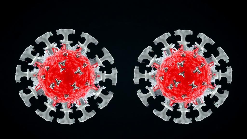
Hepatitis B virus remains one of the world’s most persistent pathogens, chronically infecting hundreds of millions of people and placing an enormous burden on global health. At the heart of the virus’s life cycle is its capsid, a protein shell assembled from dozens of copies of a single building block, the core protein. Because this assembly step is essential for viral replication, a class of experimental antiviral drugs known as capsid assembly modulators, or CAMs, has attracted intense attention. These molecules do not simply block the virus; they actively push the core protein into building malformed, noninfectious structures. A new study published in PLOS Pathogens by Liam W. Scott, Carolina Pérez-Segura, Jodi A. Hadden-Perilla, and Adam Zlotnick now reveals a subtlety in how these drugs are studied that could reshape how future CAMs are designed.
The core protein of hepatitis B virus, abbreviated HBc, normally self-assembles into icosahedral capsids, the geometrically precise containers that house the viral genome. In an infected cell, this assembly is a tightly regulated process, and disrupting it is a proven antiviral strategy. CAMs are direct-acting antivirals that induce the core protein to mis-assemble, producing empty or aberrant particles that cannot support the viral life cycle. Several CAMs have advanced into clinical development, making the structural details of how they bind to the core protein a matter of practical consequence for medicinal chemists trying to improve potency and selectivity.
Structural biologists studying CAM-core protein complexes have long relied on two different laboratory versions of the protein, and the new work systematically compares them for the first time in this context. The first, called Cp150, is the full-length form of the core protein. Cp150 forms empty icosahedral capsids that are structurally indistinguishable from the capsids found inside actual infectious virions, making it the closest available mimic of the authentic drug target. The second version, Cp149-Y132A, carries a single amino acid substitution, the Y132A mutation, which replaces a tyrosine at position 132 with an alanine. This small change has a dramatic consequence: the protein becomes defective at assembly and instead remains soluble, but it crystallizes into flat hexagonal sheets in which each hexagon resembles the quasi-sixfold vertex of an icosahedral capsid.
The Y132A variant has been a workhorse of CAM research precisely because its flat, crystalline sheets are amenable to X-ray crystallography, allowing researchers to obtain high-resolution pictures of drugs sitting in the CAM binding pocket. The implicit assumption in much of this work has been that the local environment of the binding site in the crystal sheets faithfully represents the binding site in a real capsid. The new study puts that assumption to a rigorous test by comparing structures of CAM-bound Cp150 capsids with structures of CAM-bound Cp149-Y132A crystals, asking whether the two systems tell the same story about drug recognition.
The answer, in short, is that they do not. When CAMs bind to intact capsids made of Cp150, the residues that form the CAM binding site undergo a noticeable structural adjustment, reshaping themselves to complement the geometry of the bound drug. This is a classic example of induced fit, the phenomenon in which a binding site is not a rigid lock but a malleable pocket that conforms to its ligand. In the Cp149-Y132A crystals, by contrast, the CAM sites show remarkably little structural adjustment in response to different CAMs. The pocket in the flat sheets is comparatively rigid, essentially presenting the same shape regardless of which drug is occupying it.
This difference in flexibility has a downstream consequence for which parts of the protein actually touch the drug. In the capsid structures, the array of residues that make contact with a bound CAM varies from one CAM to another, reflecting the induced-fit rearrangements of the pocket and the distinct chemistries of individual compounds. In the Cp149-Y132A crystals, that contact array remains nearly constant across different CAMs. In other words, the crystal system tends to paint a uniform picture of CAM binding, while the capsid system reveals a more dynamic and drug-dependent interaction landscape.
The implications for drug development are significant. If a CAM is optimized against the rigid binding site seen in Cp149-Y132A crystals, it may be tailored to contacts that only exist in that artificial context. In the real capsid, where the pocket flexes to embrace each ligand, the same drug might engage a different set of residues, or fail to engage the ones that matter most for potency. Conversely, the capsid-bound structures capture the induced-fit behavior that presumably operates during an actual infection, when CAM molecules encounter assembling or assembled capsids in the hepatocyte. Structures that reflect this conformational adaptability are therefore likely to be better guides for rational design.
None of this means that Cp149-Y132A crystals are without value. The variant remains a powerful tool for obtaining high-resolution structural information that is difficult or impossible to obtain from intact capsids, and the flat hexagonal sheets preserve the overall topology of the quasi-sixfold environment in which CAMs bind. The study’s point is more nuanced: the two systems provide complementary but distinctly different views of the CAM binding site, and researchers interpreting crystal structures of Cp149-Y132A complexes should be aware that the pocket they are looking at is stiffer and less responsive than its counterpart in a genuine capsid. Differences between the two systems, the authors argue, will contribute directly to future CAM design by clarifying which structural features are artifacts of the crystal system and which are intrinsic to the drug target.
The work also speaks to a broader principle in structural virology and antiviral research. Viral capsid proteins are not static scaffolds; their binding sites are shaped by the assembly state of the particle and by the partners they encounter. A protein that assembles into a closed icosahedral shell experiences constraints and interactions that a mutated, assembly-defective version in a crystal lattice does not. When those differences extend into a drug binding pocket, as they demonstrably do for the HBV core protein, the choice of structural system becomes part of the drug design problem itself. Screening and structure-based optimization campaigns that mix and match structural systems without accounting for these differences risk pursuing compounds optimized against a target that does not quite exist in the virus.
For the hepatitis B field, where CAMs represent one of the most promising new therapeutic modalities in decades, the study offers a practical roadmap. Structures of CAMs bound to Cp150 capsids capture the induced-fit flexibility of the authentic binding site and the drug-specific contact patterns that accompany it, while Cp149-Y132A crystals provide a rigid, high-resolution reference frame. Used together, and with an understanding of their differences, these two forms of the core protein can guide the design of assembly modulators that bind more effectively to the capsids that actually form during infection. The research, published in PLOS Pathogens under DOI 10.1371/journal.ppat.1014700, underscores that in the pursuit of antiviral drugs, the fine print of structural biology, down to the behavior of individual residues in a binding pocket, can determine whether a promising molecule succeeds or falls short.
Subject of Research: Structural comparison of CAM binding to assembly-active and assembly-defective forms of the hepatitis B virus capsid protein
Article Title: Assembly-active and -inactive forms of HBV capsid protein provide distinctly different binding sites for capsid assembly modulators
Article References: Scott, L. W., Pérez-Segura, C., Hadden-Perilla, J. A., & Zlotnick, A. (2026). Assembly-active and -inactive forms of HBV capsid protein provide distinctly different binding sites for capsid assembly modulators. PLOS Pathogens, 22(10), e1014700. https://doi.org/10.1371/journal.ppat.1014700
Image Credits: AI Generated
DOI: 10.1371/journal.ppat.1014700
Keywords: hepatitis B virus, HBV core protein, capsid assembly modulators, CAMs, Cp150, Cp149-Y132A, induced fit, X-ray crystallography, antiviral drug design, capsid assembly, PLOS Pathogens, structural virology
News Source: Kristina Jarvis. (October 10, 2026). Two Faces of the Hepatitis B Capsid Protein Reveal Distinct Drug Binding Sites. Scienmag.