Every surgical decision that touches a blood vessel rests on two kinds of knowledge: the static architecture of the vascular network and the dynamic behavior of blood flowing through it. The three-dimensional arrangement of vessels defines the boundaries of tissue perfusion, telling a surgeon where a tumor margin ends and where critical supply routes begin. The real-time movement of blood within those vessels reveals patency, ischemic risk, and the likelihood of functional recovery after the operation is over. A technology that could capture both dimensions, across a wide field of view and at high resolution, has long been an aspiration of optical imaging. A team at South China Normal University now reports a strategy that brings that aspiration substantially closer to clinical reality.
Writing in Light: Science & Applications, researchers led by Professor Sihua Yang of the MOE Key Laboratory of Laser Life Science and the School of Optoelectronic Science and Engineering describe PAATAM, a real-time photoacoustic angiography tracking and mapping strategy. The name compresses an ambitious goal: turning the photoacoustic vascular images themselves into the intrinsic cues that guide freehand localization and mapping, so that a clinician sweeping a handheld probe across tissue can watch a globally consistent, panoramic three-dimensional vascular map assemble on screen. The reported registration accuracy of 99.67 percent is the headline number, but the underlying engineering is what makes such accuracy possible under real scanning conditions.
To appreciate the problem PAATAM solves, it helps to understand why photoacoustic angiography has been constrained until now. The technique combines optical absorption contrast with ultrasonic detection: short laser pulses cause absorbing structures, chiefly hemoglobin in blood vessels, to emit ultrasound waves that are detected and reconstructed into images. This yields high-sensitivity, high-resolution visualization of superficial microvasculature without ionizing radiation or exogenous contrast agents. The catch is a fundamental trade-off. High-resolution photoacoustic imaging typically delivers only a small single-scan field of view, because the optical illumination and ultrasonic detection geometry that produce fine detail also limit coverage.
Freehand scanning is the obvious remedy: move the probe across the tissue and stitch the frames together. But the human hand is a poor positioning instrument, and living tissue is a hostile environment for registration algorithms. Respiration, heartbeat, tissue deformation, bleeding, and hand tremor all shift the vascular scene between acquisitions. Existing two-dimensional stitching methods can enlarge the field of view, yet they sacrifice depth information and cannot readily preserve the flow velocity and direction data that make photoacoustic imaging functionally informative. A stitched flat mosaic of vessels, however pretty, tells the surgeon little about the three-dimensional course of a vessel around a curved organ or about whether blood is still moving through it.
PAATAM’s central insight is that photoacoustic data contain two complementary families of features, and that fusing them makes localization robust where either alone would fail. The first family is vascular geometry, extracted from the three-dimensional point clouds reconstructed from continuously acquired photoacoustic data. The branching topology, vessel diameters, and spatial curvature of the network act like landmarks: distinctive structures that can be matched between successive frames to estimate how the probe has moved. The second family is absorption-related texture, drawn from intensity projection images, which encodes the signal-strength patterns of the vascular bed. Geometry anchors the map in space; texture adds discriminative detail in regions where geometry alone is ambiguous.
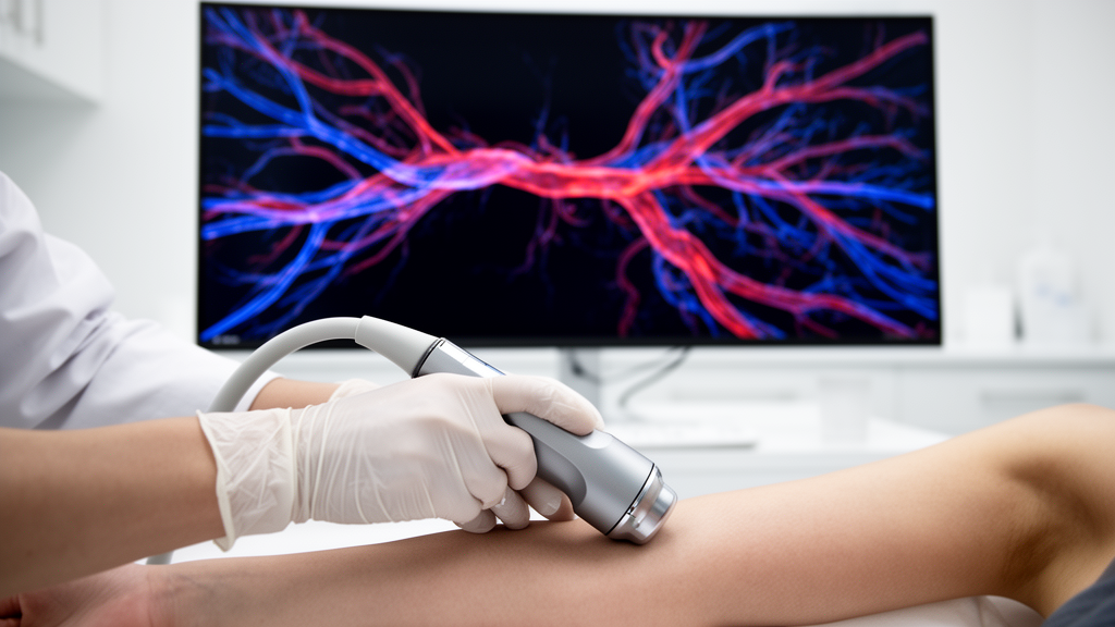
The method’s processing pipeline reflects the realities of freehand imaging. Continuously acquired photoacoustic data are reconstructed into vascular point clouds, intensity images, and depth images, which together feed real-time probe pose estimation and panoramic three-dimensional mapping. Cross-validation between the geometric and texture features guards against erroneous matches, while motion distortion correction compensates for the tissue and probe movements that would otherwise corrupt the trajectory estimate. A sliding-window factor graph optimization then refines the sequence of estimated poses, distributing errors across recent frames rather than letting them accumulate. The output is a six-degree-of-freedom estimate of handheld probe motion, sufficient to place every reconstructed vascular frame into a single coherent three-dimensional coordinate frame.
The researchers summarize the operational principle in their own words. “We establish a vascular hybrid-feature-driven localization and mapping method for freehand photoacoustic angiography,” they explain. “Continuously acquired photoacoustic data are reconstructed into vascular point clouds, intensity images, and depth images, which are used for real-time probe pose estimation and panoramic three-dimensional mapping.” The approach is conceptually a cousin of simultaneous localization and mapping, or SLAM, the family of algorithms that lets autonomous vehicles and robots build maps while tracking their own position within them. Here, however, the landmarks are living blood vessels, and the sensor is an optical-ultrasonic probe rather than a camera or lidar.
The payoff of the hybrid design is resilience against the specific failure modes of photoacoustic scanning. “By coordinating vascular geometry and signal-intensity features, the method reduces registration degradation and accumulated drift caused by tissue motion, bleeding, low-texture regions, and hand tremor,” the team notes, adding that “this enables robust three-dimensional vascular mapping on complex curved organs.” Each of those failure modes has historically been enough to derail a stitching pipeline on its own. Bleeding can obscure the very vessels being tracked; low-texture regions deprive intensity-based matching of features; tremor injects high-frequency pose noise; and curved organs such as the stomach or oral cavity violate the planar assumptions of two-dimensional mosaicking. Fusing two feature streams with cross-validation and graph optimization means that when one cue degrades, the other can carry the estimate.
Validation spanned two very different anatomical settings: human oral imaging and a rat partial gastrectomy model. In the surgical experiment, PAATAM rapidly generated a preoperative three-dimensional vascular navigation map of the gastric region, revealing the spatial relationship between the planned resection area and the vessels that needed to be preserved. Postoperative scanning then allowed the team to evaluate whether those major vessels had indeed been spared. This pre- and post-operative pairing is precisely the workflow a surgeon would want: plan the resection against a panoramic vascular map, operate, and then verify perfusion-critical structures noninvasively at the bedside.
The functional dimension of the method may prove as consequential as the structural one. Combined with photoacoustic optical-flow analysis, PAATAM captured flow velocity and direction, reflecting hemodynamic changes during vascular occlusion and reperfusion in the animal model. That means the same freehand scan that maps where the vessels are can also report whether blood is moving through them, and how fast, and in which direction. For surgical navigation, perfusion assessment, and postoperative functional evaluation, this integrated structural and functional imaging basis points toward a future in which real-time, freehand, minimally invasive photoacoustic imaging becomes a routine instrument of surgical guidance rather than a laboratory curiosity.
Subject of Research: Photoacoustic SLAM-based panoramic 3D vascular mapping for surgical navigation
Article Title: Geometry–texture dual-driven SLAM enables panoramic 3D photoacoustic vascular mapping
Article References: Geometry–texture dual-driven SLAM enables panoramic 3D photoacoustic vascular mapping. (n.d.). Original publication
Image Credits: AI Generated
DOI: Not provided
Keywords: photoacoustic angiography, SLAM, 3D vascular mapping, surgical navigation, hemodynamics, freehand scanning, point cloud registration, probe pose estimation, microvasculature, perfusion assessment, Light: Science & Applications, biomedical optics
News Source: Ophelia Keating. (October 10, 2026). Handheld probe, full picture: dual-feature SLAM builds panoramic 3D vascular maps. Scienmag.