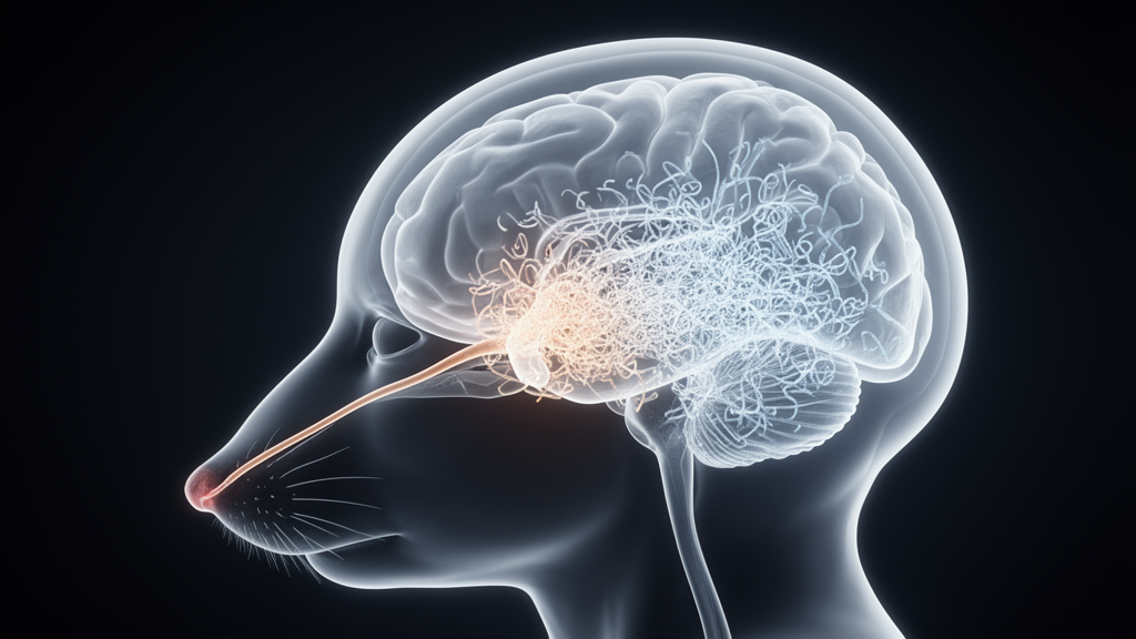
Parkinson’s disease has long been one of neuroscience’s most stubborn puzzles. Its molecular signature, the abnormal accumulation of the protein alpha-synuclein inside neurons, is easy to describe but remarkably difficult to reproduce faithfully in the laboratory. Now, a team of researchers working across India, the Czech Republic, Germany, and the United Kingdom has unveiled a mouse model that may capture the disease’s early trajectory better than most existing approaches. By delivering the mitochondrial toxin rotenone through the nose over five and a half months, the scientists produced a slow, progressive march of alpha-synuclein pathology that begins in the olfactory bulb and spreads outward to the midbrain and cortex, mirroring what many researchers call the brain-first pattern of Parkinson’s progression.
The study, published in npj Parkinson’s Disease, was led by Monika Sharma and colleagues at the National Institute of Pharmaceutical Education and Research in Ahmedabad, with senior author Amit Khairnar coordinating an international collaboration that included Tiago F. Outeiro of the University Medical Center Göttingen and Newcastle University and Irena Rektorova of Masaryk University in Brno. Their central question was deceptively simple: what happens when an environmental toxin with known links to Parkinson’s disease is introduced through the same route that many real-world exposures take, the nasal passages, and then observed over a clinically meaningful timescale?
Rotenone is a naturally occurring pesticide and a well-established inhibitor of mitochondrial complex I, the enzyme complex that cells rely on for efficient energy production. Chronic systemic rotenone exposure has been used for years to induce parkinsonian neurodegeneration in rodents, but those models typically flood the entire body with the toxin, making it hard to determine where the disease process actually starts. The intranasal route changes that calculus. By administering rotenone directly into the nose of C57BL/6 male mice, the researchers exploited the anatomical connections between the olfactory epithelium, the olfactory bulb, and deeper brain structures, creating a plausible entry point for a pathological cascade that begins in the brain rather than the gut.
This distinction matters because the field is divided over the direction in which Parkinson’s pathology travels. The influential Braak staging hypothesis proposes that in many patients, the disease begins in the enteric nervous system or olfactory structures and ascends toward the substantia nigra, the midbrain region whose dopamine-producing neurons die in Parkinson’s disease. A brain-first variant, in which pathology originates in the olfactory bulb and spreads caudally, has been proposed for a subset of patients who experience early loss of smell without prominent gastrointestinal symptoms. Animal models that faithfully reproduce one or the other pattern have been scarce, which is precisely the gap this study set out to fill.
The experimental design was longitudinal and deliberately unhurried. The mice received intranasal rotenone for a total of five and a half months, and the researchers examined them at three time points: three months, four months, and five and a half months after exposure began. At each stage they combined behavioral testing with molecular and cellular analysis. Olfactory function was assessed to detect the earliest sensory deficits, while motor tests tracked the emergence of the movement impairments that define clinical Parkinson’s disease. In parallel, the team used immunoblotting and immunofluorescence to map the accumulation of alpha-synuclein, the activation of glial cells, and the survival of dopaminergic neurons across multiple brain regions.
The results unfolded in a strikingly time-dependent sequence. Alpha-synuclein accumulation appeared first in the olfactory bulb and then, as the months passed, was detected progressively in more distant regions, including the midbrain and the cortex. This spatial spread was not an isolated molecular event. It was accompanied by measurable atrophy of the olfactory bulb itself, a growing burden of neuroinflammation marked by activated glial cells, and the gradual degeneration of dopaminergic neurons. Behavioral performance deteriorated in step with the pathology, with olfactory deficits preceding the motor impairments that would ordinarily dominate a parkinsonian phenotype.
One of the most intriguing findings to emerge from the study is a link between distinct patterns of astrocyte activation and dopaminergic activity. Astrocytes, the star-shaped support cells of the central nervous system, are increasingly recognized as active participants in neurodegeneration rather than passive bystanders. Reactive astrocytes can adopt different functional states, some of which are protective and others inflammatory. By establishing a correlation between a specific astrocyte activation profile and the health of dopaminergic neurons, the study adds a cellular dimension to the model that could prove valuable for drug screening. If astrocyte states can be modulated to protect vulnerable neurons, this model offers a platform for testing such interventions in a setting where the pathological timeline is known and predictable.
Technically, the model’s strengths lie in its chronicity and its anatomical fidelity. Many rodent models of Parkinson’s disease rely on acute toxin administration or on the injection of preformed alpha-synuclein fibrils directly into the brain, approaches that produce robust pathology but bypass the question of how the disease might begin. The intranasal rotenone model, by contrast, starts the cascade at a peripheral sensory interface and lets it develop over months, closer to the tempo of human disease. The progressive nature of the pathology also creates multiple therapeutic windows: researchers can intervene early, when alpha-synuclein is confined to the olfactory bulb, or later, when it has reached the midbrain, and compare outcomes. That kind of staged testing is essential for evaluating disease-modifying therapies, which are intended to slow or halt progression rather than simply relieve symptoms.
The model also reinforces the growing appreciation of environmental contributions to Parkinson’s disease. Epidemiological studies have repeatedly associated pesticide exposure, including rotenone, with elevated Parkinson’s risk, and the nose-to-brain pathway has been proposed as a plausible route by which inhaled toxins could seed pathology in the olfactory system. By demonstrating that chronic intranasal exposure is sufficient to drive a brain-first pattern of alpha-synuclein spread, neuroinflammation, and dopaminergic degeneration, the study provides experimental support for a mechanism that has been largely inferential. It does not prove that human Parkinson’s disease is caused by inhaled pesticides, but it shows that such an exposure route can generate the characteristic pathological sequence in a mammalian brain.
The authors conclude that their chronic and progressive mouse model mimics the brain-first type of progression seen in some Parkinson’s patients, opening the possibility of testing potential disease-modifying interventions under conditions that resemble the earliest stages of the human illness. For a field in which most therapies address symptoms after substantial neuronal loss has already occurred, a model that captures the disease’s opening moves could shift the focus toward prevention and early intervention. The work was supported by the National Institute of Pharmaceutical Education and Research seed fund, the Ramalingaswami Fellowship from the Department of Biotechnology, India, and imaging infrastructure funded through Czech-BioImaging and European Union programs, reflecting the increasingly international and interdisciplinary character of Parkinson’s research. As the model is adopted and refined by other laboratories, its true value will be measured by whether therapies validated within it can finally change the trajectory of a disease that currently only moves in one direction.
Subject of Research: A chronic intranasal rotenone mouse model of brain-first alpha-synuclein pathology progression in Parkinson's disease
Article Title: A ‘brain-first’ mouse model of progressive alpha-synuclein pathology via intranasal rotenone administration
Article References: Sharma, M., Sharma, N., Soni, J., Uttarkar, M., Ruda-Kucerova, J., F. Outeiro, T., Rektorova, I., & Khairnar, A. (2026). A ‘brain-first’ mouse model of progressive alpha-synuclein pathology via intranasal rotenone administration. npj Parkinson's Disease. https://doi.org/10.1038/s41531-026-01593-4
Image Credits: AI Generated
DOI: 10.1038/s41531-026-01593-4
Keywords: Parkinson's disease, alpha-synuclein, rotenone, intranasal administration, olfactory bulb, brain-first model, dopaminergic neurodegeneration, neuroinflammation, astrocyte activation, mouse model, environmental toxins, mitochondrial complex I
News Source: Cassandra Pierce. (October 9, 2026). Intranasal Rotenone Model Recreates Brain-First Spread of Parkinson’s Pathology in Mice. Scienmag.