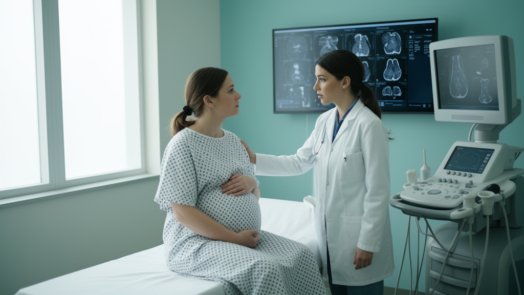
A rare and aggressive form of ovarian cancer has been diagnosed in a 35-year-old woman at just ten weeks of pregnancy, in a case that underscores how difficult it can be to distinguish a serious malignancy from the ordinary discomforts of early gestation. The case, published in Volume 13 of the journal Oncoscience on September 24, 2026, describes high-grade serous carcinoma of the ovary presenting as a complex cystic mass during the first trimester, a combination so uncommon that clinicians have few established guidelines to follow. The report was led by first author Chanderdeep Sharma from the All India Institute of Medical Sciences in Bilaspur, Himachal Pradesh, India, with corresponding author Shri Ram Rundla also affiliated with the same institution. Together, the clinical team documented not only the tumor itself but the entire chain of diagnostic reasoning, imaging findings, surgical decisions and follow-up care that such an exceptional situation demands.
Ovarian cancer complicating pregnancy is uncommon by any measure, with an estimated incidence ranging from one in 12,000 to one in 47,000 pregnancies. Within that already narrow category, high-grade serous carcinoma occupies a particularly concerning niche. It is an aggressive epithelial ovarian cancer, and its diagnosis during early gestation is exceptionally rare. The difficulty begins with symptoms. Abdominal bloating, discomfort and nausea are hallmark complaints of normal early pregnancy, yet they are also the vague presenting signs of ovarian malignancy. This overlap means that a growing tumor can be mistaken for pregnancy-related changes, delaying recognition of a potentially life-threatening disease at a stage when both maternal health and the ongoing pregnancy hang in the balance.
In the reported case, the patient arrived with lower abdominal pain and progressive abdominal distension that had persisted for approximately two weeks. She had previously delivered two children and reported no personal or family history of malignancy, factors that might otherwise have lowered suspicion. Clinical examination, however, identified a palpable pelvic mass, prompting further investigation. Ultrasonography confirmed a viable pregnancy but also revealed a large mass measuring 10 by 13 centimeters in the left adnexa, the anatomical region surrounding the ovary. The mass was complex, containing both solid and cystic components separated by internal septations, and it was accompanied by ascites, an abnormal accumulation of fluid within the abdominal cavity, as well as omental nodularity. Each of these features independently raised concern; together, they painted a picture strongly suggestive of ovarian malignancy.
Laboratory testing added another layer of complexity. The patient’s cancer antigen 125, or CA-125, level was elevated at 332 units per milliliter. CA-125 is a widely used tumor marker in ovarian cancer management, but it carries a well-known limitation in pregnancy: it may rise naturally during early gestation and cannot independently establish a diagnosis of ovarian cancer. In this case, the elevated marker could not be interpreted in isolation, but when considered alongside the suspicious imaging findings, it strengthened the argument for further evaluation. This interplay between a marker that is unreliable in pregnancy and imaging features that are difficult to interpret in a gravid abdomen illustrates precisely why early-stage diagnosis of ovarian malignancy during gestation remains such a formidable clinical challenge.
Given the concern for advanced malignancy, the clinical team turned to contrast-enhanced computed tomography, a decision that required careful weighing of potential maternal benefit against fetal risk from ionizing radiation. The patient was counseled about both dimensions, and informed consent was obtained before the scan proceeded. The CT confirmed the complex adnexal mass, the ascites and peritoneal nodularity, findings suggesting that the disease might have spread beyond the ovary. The step from ultrasound to CT in a pregnant patient is not taken lightly, and the case demonstrates how, when suspicion of advanced cancer is high enough, the potential threat to the mother can justify imaging that would otherwise be deferred during gestation.
The case was then reviewed by a multidisciplinary team that brought together gynecologic oncology, radiology and anesthesia specialists. The clinicians weighed the suspected advanced stage of the disease, the potential threat to maternal health, and the implications of any treatment for the ongoing pregnancy. After detailed counselling and discussion of the available options, the patient, who had completed her reproductive plans, chose definitive surgical management. This decision-making process, documented step by step in the report, reflects the reality that no standardized pathway exists for such rare presentations. Each element, from the timing of intervention to the extent of surgery, had to be individualized on the basis of the specific clinical circumstances and the patient’s own values and preferences.
During staging laparotomy, surgeons identified a large solid-cystic tumor arising from the left ovary, along with ascites and multiple nodules involving the omentum and the peritoneal lining. Intraoperative frozen-section analysis, which allows rapid microscopic assessment of tissue during surgery, suggested epithelial ovarian carcinoma. The surgical team subsequently performed a total abdominal hysterectomy, bilateral salpingo-oophorectomy and infracolic omentectomy, removing the uterus, both ovaries and fallopian tubes, and part of the omentum. Final histopathological examination confirmed high-grade serous carcinoma, classified as FIGO stage IIIB. Under the microscope, the tumor displayed the characteristic malignant features of this entity, including marked nuclear abnormalities and frequent atypical cell divisions. Immunohistochemical testing demonstrated strong nuclear positivity for WT1, a marker that supported the diagnosis of serous ovarian carcinoma and helped distinguish it from other epithelial subtypes.
Following an uneventful postoperative recovery, the patient received adjuvant chemotherapy with paclitaxel and carboplatin, a standard regimen for this tumor type. At the reported one-year follow-up, she remained disease-free and continued regular surveillance. The case also raises important considerations about treatment timing during pregnancy. Surgical intervention for suspected ovarian malignancy during gestation is generally postponed until the second trimester when feasible, a window considered safest for the developing fetus. However, when imaging suggests advanced malignancy and maternal health may be at risk, intervention during the first trimester may become necessary. The authors emphasize that such decisions require individualized assessment, informed consent and coordination among the relevant specialists, rather than rigid adherence to any single timeline.
The report also turns to the potential role of hereditary cancer susceptibility. High-grade serous ovarian carcinoma is frequently associated with alterations involving the TP53 gene, while inherited mutations in BRCA1 and BRCA2 can influence both cancer risk and treatment options. Because of the patient’s relatively young age at diagnosis, genetic counselling and BRCA testing were recommended. In this instance, however, testing could not be performed because of logistical constraints, leaving her genetic status unknown. This gap highlights a practical barrier that persists in many health care settings: even when genetic evaluation is clearly indicated, access and resources may determine whether it actually takes place, with potential consequences for the patient’s relatives and for future treatment decisions.
As a single-patient case report, the findings cannot establish how frequently high-grade serous ovarian carcinoma occurs during pregnancy, nor can they determine which treatment approach produces the best outcomes across different clinical circumstances. Nevertheless, the case provides a concrete example of the diagnostic and therapeutic decisions involved when a potentially advanced ovarian malignancy is discovered during early gestation. The authors summarize the central lesson in their own words: early imaging, appropriate surgical planning, and multidisciplinary decision-making are essential for optimal maternal outcomes. Overall, the report underscores the importance of thoroughly evaluating suspicious adnexal masses during pregnancy, particularly when complex imaging features, ascites and elevated tumor markers converge to raise concern for malignancy. In these uncommon and challenging clinical situations, early recognition, coordinated specialist care and individualized treatment planning may make the decisive difference in protecting maternal health while navigating the profound stakes of early pregnancy.
Subject of Research: High-grade serous ovarian carcinoma diagnosed during early pregnancy
Article Title: Rare ovarian cancer diagnosed during early pregnancy highlights diagnostic challenges
Article References: Rare ovarian cancer diagnosed during early pregnancy highlights diagnostic challenges. (n.d.). Original publication
Image Credits: AI Generated
DOI: Not provided
Keywords: ovarian cancer, high-grade serous carcinoma, pregnancy, adnexal mass, CA-125, WT1, staging laparotomy, BRCA1, BRCA2, chemotherapy, multidisciplinary care, Oncoscience
News Source: Ophelia Keating. (October 9, 2026). Aggressive Ovarian Cancer Found at 10 Weeks of Pregnancy Exposes Diagnostic Dilemmas. Scienmag.