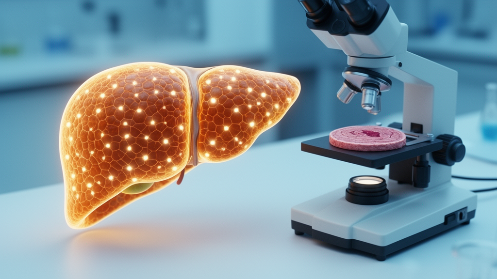
How does an organ build itself from a handful of cells into a functioning, centimeter-scale structure? For decades, biologists could only answer that question in fragments, sampling thin slices of tissue and inferring the whole from the parts. Now a team led by Chen-Hui Chen at Academia Sinica in Taiwan has done something that sounds almost impossible: they have counted and mapped every single hepatocyte in an intact adult vertebrate liver, tracking how the organ’s cellular population changes from larval life through full adulthood. The study, published in PLOS Biology, introduces a workflow called whole adult-organ expansion microscopy, or WAO-ExM, and uses it to rewrite the story of how the zebrafish liver grows.
The technical hurdle the researchers faced is deceptively simple to state. Adult organs are large, measured in millimeters to centimeters, while the cells that compose them are measured in micrometers. Conventional light microscopes cannot resolve every nucleus throughout a thick, intact organ, and sectioning the organ into slices destroys the spatial relationships that matter for understanding growth. Expansion microscopy, first developed in the mid-2010s, offered a way out by physically swelling tissue samples so that their features spread apart and become resolvable under standard optics. But applying the technique to an entire adult organ, rather than a thin slice, required a substantial engineering effort to make the method uniform, robust, and compatible with fluorescent reporters that label specific cell types.
The team’s solution was an integrated pipeline that combines tissue clearing, gel embedding, isotropic expansion, and high-speed light-sheet fluorescence imaging. They worked with transgenic zebrafish whose hepatocyte nuclei carry fluorescent tags, so every liver cell could be individually identified in the final images. After expansion and imaging, computational analysis stitched the data into a complete three-dimensional atlas of the liver, in which each hepatocyte could be located, counted, and measured. The result is a genuine whole-organ census: an intact adult zebrafish liver, spanning roughly five millimeters in thickness, was found to contain an average of 1,265,206 hepatocytes. That number, obtained by direct observation rather than extrapolation, sets a new benchmark for quantitative organ biology.
With the census in hand, the researchers turned to the central question of growth dynamics. They imaged livers at multiple stages across the post-embryonic period, from juvenile fish through fully mature adults, and charted how hepatocyte numbers changed over time. The pattern they found was strikingly non-linear. Rather than growing steadily in proportion to the body, the liver’s cell population increased by a remarkable 538-fold, and much of that increase was packed into a temporally concentrated burst of proliferation. In other words, the liver does not simply scale up with the rest of the animal; it follows its own growth schedule, with an intense phase of cell addition that is decoupled from overall body growth.
This decoupling is more than a curiosity. It suggests that organs possess intrinsic growth programs that operate on their own timelines, potentially governed by internal cues such as local signaling gradients, mechanical stress, or metabolic demand rather than by systemic growth factors alone. For developmental biologists, the finding reframes post-embryonic organ growth as an active, staged process rather than a passive consequence of the animal getting bigger. It also raises new questions about how a growing liver maintains its architecture, its blood supply, and its bile duct network while its cell population expands by more than two orders of magnitude in a relatively short window.
Counting cells, however, only tells part of the story. The deeper question is which cells are responsible for the growth. Are new hepatocytes generated broadly, with most existing cells dividing a little, or are they the descendants of a small subset of progenitors that divide a lot? To find out, the team integrated lineage tracing with their expansion microscopy workflow. In lineage tracing, a small number of cells are permanently labeled with a heritable fluorescent marker, and all of their descendants carry that same label. By combining this approach with whole-organ imaging, the researchers could trace clone territories, the spatial footprints of individual founding cells, across the entire adult liver.
The answer was unambiguous: liver growth is driven by a few hepatocytes undergoing drastic clonal expansion at the whole-organ scale. Instead of a democratic process in which every cell contributes equally, the growing liver is dominated by a small number of lineages whose progeny multiply extensively and come to occupy large territories within the organ. This clonal organization echoes principles seen in other self-renewing tissues, such as the intestinal epithelium and the blood system, where a limited pool of highly proliferative cells sustains tissue mass. Seeing the same logic play out across an entire intact vertebrate organ, rather than in a two-dimensional section, gives the observation a new level of rigor and visual force.
The clonal picture also has implications for how we think about liver disease and regeneration. The mammalian liver is famous for its ability to regrow after injury, and hepatocellular carcinoma is often discussed in terms of clonal expansion of mutated cells. If normal liver growth in zebrafish is naturally driven by a few dominant clones, then the boundary between physiological growth and pathological expansion may be governed by regulatory mechanisms that keep clonal proliferation in check. Understanding those mechanisms in a transparent, genetically tractable vertebrate could illuminate what goes wrong when clonal control fails in human disease.
The researchers did not stop at cell numbers and clones. They also asked how the liver’s three-dimensional shape emerges during growth, and whether shape and cell number are controlled by the same or different mechanisms. Their target was the extracellular matrix, specifically laminins, a family of proteins that form a structural scaffold around tissues and are known to influence cell behavior. When the team disrupted laminin function, they observed a striking dissociation: the liver’s overall shaping was perturbed even as total cell number increases continued. This decoupling suggests that tissue architecture and cell proliferation are regulated independently, with the extracellular matrix acting as an architectural control layer that is at least partly separate from the growth control layer. For bioengineers trying to grow organs in the lab, the lesson is that building the right structure requires more than making enough cells; the scaffold matters on its own terms.
Perhaps the most exciting aspect of the study is its generality. The authors showed that WAO-ExM is not limited to healthy livers. They applied the workflow to diseased livers, where it could resolve pathological changes at single-cell resolution across the whole organ, and to other adult zebrafish organs, including the heart and the pancreas. In each case, the platform delivered the same promise: a complete, single-cell-resolved view of an intact adult vertebrate organ. Because zebrafish are transparent, genetically manipulable, and widely used in biomedical research, the method is likely to spread quickly through labs studying regeneration, cancer, diabetes, and congenital heart disease. The technique bridges micrometer-scale cell behaviors with centimeter-scale organ growth, closing a persistent gap in biology between what we can see in a dish and what happens in a living body. For a field that has long had to choose between seeing everything and seeing clearly, whole-organ expansion microscopy offers, for the first time, both at once.
Subject of Research: Single-cell mapping of growth dynamics and clonal organization in the developing and adult zebrafish liver
Article Title: Whole-organ single-cell mapping defines growth dynamics and clonal organization in developing and adult zebrafish livers
Article References: Roan, H.-Y., Tian, X., Chu, W.-C., Lee, C.-M., Chen, H., Santoso, F., Wang, C.-H., Liu, Y.-H., Kumar, U., Hsiao, H.-Y., Yuh, C.-H., Chen, B.-C., & Chen, C.-H. (2026). Whole-organ single-cell mapping defines growth dynamics and clonal organization in developing and adult zebrafish livers. PLOS Biology, 24(9), e3004007. https://doi.org/10.1371/journal.pbio.3004007
Image Credits: AI Generated
DOI: 10.1371/journal.pbio.3004007
Keywords: zebrafish, liver, expansion microscopy, hepatocytes, clonal expansion, organ growth, lineage tracing, extracellular matrix, laminin, single-cell imaging, PLOS Biology, regeneration
News Source: Drew Townsend. (October 9, 2026). Every Liver Cell Counted: Whole-Organ Microscopy Reveals How Organs Grow. Scienmag.