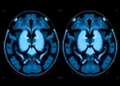
Traumatic brain injury kills and disables millions of people every year, with an estimated 20.8 million new cases annually worldwide, and a growing body of evidence suggests that one of the most decisive factors in survival may be hiding in plain sight inside the blood. A new viewpoint published in the journal Neurocritical Care by Manuel Quintana Diaz of La Paz University Hospital in Madrid and Daniel Agustin Godoy of the Meditech Foundation in Cali, Colombia, argues that the field of neurocritical care has been measuring the wrong thing. For decades, clinicians have judged the platelet system of injured brains by a single number: the platelet count. The authors contend that this binary approach misses a dynamic, biologically rich phenomenon they call the hemostatic phenotype, and that what cannot be seen on a standard count may be exactly what determines whether a patient’s intracranial bleed smolders quietly or expands catastrophically.
The core of the argument is deceptively simple. A platelet count tells you how many platelets are circulating, but says nothing about whether they work. Platelets contribute to hemostasis through adhesion, activation, granule release, aggregation, and support of thrombin generation, and none of these functions is captured by a routine count. Multiple studies have now documented profound platelet dysfunction in patients with traumatic brain injury even when their counts sit comfortably within the normal range. The authors define this dysfunction as a measurable impairment in agonist-induced aggregation, in the responsiveness of specific receptor pathways, or in the platelet’s contribution to whole-blood clot strength, occurring disproportionately to or entirely in the absence of thrombocytopenia. In other words, a patient can have plenty of platelets and still have platelets that fail.
The biological explanation lies in the unique nature of brain injury. When neural tissue is disrupted, it releases tissue factor, damage-associated molecular patterns, microparticles, and lipid mediators into the circulation, triggering thrombin generation, platelet activation, and ultimately receptor desensitization. The result is a paradoxical hemostatic state the authors describe as activation exhaustion: early procoagulant activation coexists with downstream hyporesponsiveness. Platelet activation and platelet dysfunction, far from being mutually exclusive, appear to be overlapping temporal phases of the same post-injury response. This may explain a frustrating clinical pattern in which patients with traumatic brain injury suffer early hemorrhagic progression of their intracranial lesions and later develop thrombotic complications, two seemingly opposite outcomes arising from the same underlying biology.
The strongest mechanistic evidence comes from a study by Castellino and colleagues, who examined 70 patients with isolated head injury, carefully excluding hemorrhagic shock, hypotension, and antiplatelet medication use. In severe traumatic brain injury, they found adenosine diphosphate, or ADP, pathway inhibition of 93.1 percent, with an interquartile range of 44.8 to 98.3 percent, compared with just 15.5 percent in controls, a difference that was highly statistically significant, and the arachidonic acid pathway was involved concurrently. A parallel rat model confirmed ADP inhibition of 77.6 percent, plus or minus 6.7 percent, just 15 minutes after injury. These findings establish that brain injury alone, independent of systemic hemodynamic compromise, generates profound receptor-level platelet dysfunction, a mechanism distinct from pharmacological blockade and from inhibition driven by circulating mediators.
Current clinical guidelines recommend maintaining platelet counts above 50 billion per liter in active intracranial hemorrhage and above 100 billion per liter around surgery. Those thresholds remain essential, the authors stress, but they address only quantity. A patient whose count is well above both limits may still harbor significant dysfunction at the level of the ADP/P2Y12 or arachidonic acid/cyclooxygenase-1 receptor pathways. The authors also warn against two opposite errors: equating thrombocytopenia with platelet failure, and treating any detected dysfunction as an automatic indication for transfusion. Their framework rests on three clinical questions: which phenotype is present, is it clinically meaningful, and can correcting it actually improve outcomes?
Detecting these phenotypes requires functional testing, and here the landscape is complicated. Standard coagulation tests, including prothrombin time, international normalized ratio, and fibrinogen measurement, are essentially blind to platelet function. Viscoelastic assays such as thromboelastography and rotational thromboelastometry assess whole-blood clot kinetics and strength, and platelet mapping modules add agonist-specific response data. Other platforms, including Multiplate, VerifyNow, and light transmission aggregometry, measure different biological parameters and cannot be directly compared with one another. Cross-platform equivalence in traumatic brain injury has not been established, and the most frequently cited working thresholds, ADP or arachidonic acid inhibition greater than 60 percent on thromboelastography with platelet mapping, were never prospectively validated. They represent observational approximations, not clinical standards, and no universally accepted intervention cutoffs exist for any platform.
The therapeutic evidence is strikingly discordant, and the authors argue that the discordance follows a pattern rather than being random. On the positive side, one retrospective series found that goal-directed platelet transfusion guided by platelet mapping in severe traumatic brain injury was associated with lower mortality, and a platelet mapping algorithm reduced transfusion exposure without apparent harm in patients exposed to antiplatelet drugs. Against this, multiple studies found no benefit and potential harm from empirical platelet transfusion or desmopressin in less severely injured populations or when treatment was not guided by functional testing. The positive studies were conducted in severe injury with documented profound dysfunction and function-guided decisions; the negative studies applied empirical treatment in milder populations. The lesson, the authors conclude, is that intervention should be phenotype-guided and risk-stratified, not routine and empirical.
The risks of getting this wrong are not trivial. Platelet transfusion carries the danger of transfusion-associated circulatory overload, transfusion-related acute lung injury, alloimmunization, and prothrombotic complications, while desmopressin, typically dosed at 0.3 to 0.4 micrograms per kilogram intravenously in structured protocols, can cause hyponatremia, fluid retention, and thrombosis. The authors frame their recommendation through the lens of patient blood management: the question is not whether platelets are good or bad, but whether a clinically relevant, modifiable phenotype exists in which intervention is likely to change meaningful outcomes. They propose a five-category taxonomy matching platelet-related phenotypes to clinical context, dominant mechanism, and therapeutic implication, while candidly noting that this construct is expert-synthesized and has not been prospectively validated as a diagnostic classification system.
The practical approach they sketch is deliberately cautious. Platelet function testing is most defensible in moderate-to-severe injury, traumatic intracranial hemorrhage, radiological progression, planned neurosurgery or invasive neuromonitoring, known preinjury antiplatelet exposure, systemic polytrauma with coagulopathy, or unexplained bleeding despite a normal count. When function is preserved, empirical reversal should generally be avoided. When dysfunction is documented in high-risk patients requiring surgery or invasive monitoring, desmopressin or platelet transfusion may be considered according to phenotype, antiplatelet exposure, count, and procedural urgency, with post-intervention reassessment by clinical status, imaging, and repeat functional testing. Centers without access to viscoelastic platforms can fall back on clinical risk stratification using the Glasgow Coma Scale, lesion subtype, antiplatelet exposure, and procedural urgency, consistent with American College of Surgeons guidance. In emergent surgical scenarios, urgency simply overrides testing; in nonemergent settings, a 30-to-60-minute window permits targeted hemostatic preparation.
The authors are explicit about the limits of their case. Most clinical evidence is observational, retrospective, or single-center; interassay discordance remains a fundamental barrier; dysfunction rarely occurs in isolation and overlaps with fibrinogen abnormalities, acidosis, hypothermia, hypocalcemia, and hemodilution; the dynamic evolution of dysfunction over the first 72 hours is poorly characterized; and findings from moderate-to-severe cohorts cannot be extrapolated to mild injury. Many studies report laboratory correction without demonstrating improved patient outcomes. What they call for is a new generation of prospective, multicenter trials to determine which assay best identifies clinically meaningful dysfunction, which threshold predicts hemorrhagic progression, and whether phenotype-guided correction improves neurological outcomes rather than merely normalizing laboratory values. In neurocritical care, they write, what is hidden beyond the platelet count may be clinically decisive, and the next step is not simply to transfuse more platelets but to understand which platelet phenotype matters, when it matters, and how it can be safely corrected.
Subject of Research: Platelet dysfunction as a hemostatic phenotype in traumatic brain injury coagulopathy
Article Title: Rethinking Platelet Dysfunction in Traumatic Brain Injury: From Platelet Count to Hemostatic Phenotype
Article References: Quintana Diaz, M., & Godoy, D. A. (2026). Rethinking Platelet Dysfunction in Traumatic Brain Injury: From Platelet Count to Hemostatic Phenotype. Neurocritical Care. https://doi.org/10.1007/s12028-026-02670-5
Image Credits: AI Generated
DOI: 10.1007/s12028-026-02670-5
Keywords: traumatic brain injury, platelet dysfunction, coagulopathy, hemostatic phenotype, thromboelastography, platelet mapping, platelet transfusion, desmopressin, intracranial hemorrhage, neurocritical care, patient blood management, P2Y12 pathway
News Source: Cassandra Pierce. (October 6, 2026). Hidden Platelet Failure May Decide Who Survives Traumatic Brain Injury. Scienmag.