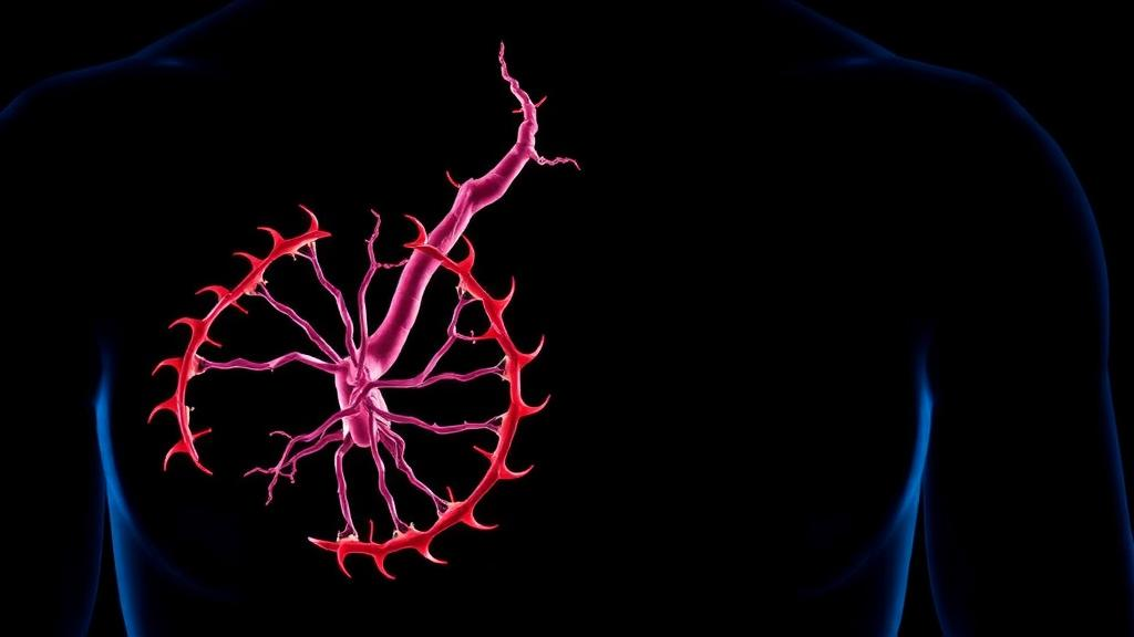
Abdominal aortic aneurysm is one of the most treacherous conditions in cardiovascular medicine. The abdominal aorta silently balloons over months or years, producing no symptoms until the vessel wall finally gives way. When rupture occurs, mortality can reach 80 to 90 percent, and many patients die before they ever reach a hospital. Despite decades of progress in imaging and surgical repair, the molecular events that drive the aneurysmal wall toward dilation and failure remain poorly understood, leaving clinicians without effective drugs to slow or prevent the disease. A new study published in the journal iScience now points to an unexpected culprit: a protein best known for helping to assemble the mitochondrial respiratory chain.
The research team, led by investigators studying the genetic landscape of aneurysm tissue, began with a large-scale bioinformatics sweep. They pooled gene expression data from three public datasets of human aortic tissue, comparing aneurysm samples with healthy controls. Principal component analysis showed that the two groups separated cleanly by their gene expression profiles, and differential expression analysis identified 617 genes that were upregulated and 711 genes that were downregulated in aneurysmal tissue. To make sense of this flood of data, the researchers applied weighted gene co-expression network analysis, which clusters genes that move together across samples. Of 32 modules identified, one dark-gray module containing 1,659 genes showed the strongest correlation with aneurysm status and became the focus of further analysis.
The team then intersected this module with two curated gene sets that they suspected might matter in aneurysm biology. The first contained mitochondrial-related genes drawn from the Human MitoCarta3.0 database, capturing proteins involved in oxidative phosphorylation and mitochondrial maintenance. The second captured genes linked to PANoptosis, a recently described form of programmed cell death that combines features of apoptosis, pyroptosis, and necroptosis and is tightly intertwined with inflammatory signaling. This cross-referencing yielded 11 mitochondrial differentially expressed genes and 10 PANoptosis-related differentially expressed genes associated with the disease. Three machine-learning algorithms, LASSO logistic regression, support vector machine recursive feature elimination, and random forest, were then used in parallel to whittle these lists down to a handful of candidate key genes, a strategy designed to reduce the bias that any single algorithm can introduce.
Seven genes survived the full gauntlet of filters: MTHFD2, NDUFAF2, ACADVL, and GLRX5 on the mitochondrial side, and CCT5, RPS27A, and PELI1 on the PANoptosis side. When the researchers measured these candidates in the laboratory, one stood out dramatically. NDUFAF2, short for NADH dehydrogenase ubiquinone 1 alpha subcomplex assembly factor 2, showed the largest expression increase during aneurysm development. The elevation was confirmed at the protein level by immunofluorescence and western blotting in cells treated with angiotensin II, a hormone-like peptide long used to model aneurysm biology, and again in the aortic tissue of aneurysm mice by quantitative PCR and immunohistochemistry. NDUFAF2 is not an obscure player: it serves as an assembly factor for mitochondrial complex I, the first and largest enzyme of the respiratory chain, and mutations in its gene are known to cause severe mitochondrial disease in humans.
To test whether NDUFAF2 was merely a bystander or an active participant, the researchers silenced it with small interfering RNA in mouse vascular smooth muscle cells exposed to angiotensin II. The results were striking. Cells lacking NDUFAF2 proliferated at a significantly higher rate and died at a significantly lower rate than controls. Flow cytometry revealed a sharp drop in apoptosis, and the molecular signature of cell death shifted accordingly: the anti-apoptotic protein Bcl-2 rose, while the pro-apoptotic proteins Bax and cleaved caspase-3 fell. Angiotensin II also crippled the migratory capacity of smooth muscle cells, an effect that NDUFAF2 knockdown largely reversed. Because the loss and dysfunction of vascular smooth muscle cells is considered a central event in aneurysm formation, these findings positioned NDUFAF2 as a plausible mechanistic link between mitochondrial stress and wall weakening.
The next question was how NDUFAF2 exerts this influence, and the answer lay in the mitochondria themselves. MitoTracker staining showed that angiotensin II drastically reduced mitochondrial mass in smooth muscle cells, and transmission electron microscopy revealed swollen organelles with fragmented cristae, the folded inner membranes where energy production takes place. Silencing NDUFAF2 reversed both changes. The protein also rebalanced mitochondrial dynamics, the constant process of fission and fusion that keeps these organelles healthy. Angiotensin II increased DRP1, a fission protein whose overactivity fragments mitochondria, while suppressing OPA1, a fusion protein of the inner membrane that maintains cristae structure and cell survival. NDUFAF2 knockdown lowered DRP1 and restored OPA1, and overexpressing OPA1 in rescue experiments further protected mitochondrial mass, membrane potential, and respiratory function, confirming that OPA1 sits downstream in this protective pathway.
Measurements of mitochondrial respiration told a consistent story. Using a Seahorse analyzer to track the oxygen consumption rate, the researchers found that angiotensin II depressed both basal and maximal respiration, indicating impaired oxidative phosphorylation, and that NDUFAF2 knockdown partially restored these values. Damaged mitochondria are a major source of reactive oxygen species, and MitoSOX staining confirmed that silencing NDUFAF2 significantly reduced mitochondrial superoxide production. This matters because excess mitochondrial ROS does more than damage lipids, proteins, and DNA; it can activate NLRP3 inflammasomes, fuel the release of pro-inflammatory cytokines such as interleukin-1 beta, and amplify the chronic inflammation and matrix degradation that characterize the aneurysmal wall. The same protective pattern held in human aortic smooth muscle cells, where NDUFAF2 knockdown reduced ROS, restored DRP1 and OPA1 balance, improved respiration, and lowered apoptotic rates.
The decisive test came in living animals. The researchers induced aneurysms in male C57BL/6J mice by applying calcium chloride solution to the abdominal aorta, a widely used model of rapid aneurysmal dilation, and delivered anti-NDUFAF2 siRNA by tail vein injection. Four weeks later, the aortas of treated mice were measurably healthier. Abdominal aortic diameters were significantly smaller than in untreated aneurysm controls, hematoxylin and eosin staining showed improved tissue architecture, and elastic fiber staining revealed markedly less degradation of elastin, the resilient protein that gives the aortic wall its recoil. TUNEL assays confirmed that cell death in the vessel wall had dropped, and western blots of aortic tissue showed the same molecular shifts seen in culture: less DRP1, Bax, and cleaved caspase-3, and more OPA1 and Bcl-2. Immunofluorescence for TOM20, a mitochondrial outer membrane marker, and alpha-SMA, a smooth muscle marker, showed that both mitochondrial content and smooth muscle cell preservation were restored, as were the electron transport chain subunits SDHA and SDHB and the contractile marker SM22alpha.
The study’s authors are careful about its boundaries. Their in vivo work relied on a single aneurysm model and only male animals, so the findings will need validation in angiotensin II perfusion models and in both sexes. The precise mechanism by which NDUFAF2 regulates mitochondrial function also remains to be worked out in depth. Even so, the work delivers a conceptually important message: a complex I assembly factor, previously studied mainly in the context of neurodegenerative mitochondrial disease and cancer prognosis, acts as a driver of vascular smooth muscle cell death in aneurysm disease. The finding resonates with earlier reports that mitochondrial NAD+ deficiency impairs type III collagen turnover and triggers aortic aneurysms, tying mitochondrial metabolism to the structural integrity of the vessel wall. If future work confirms that pharmacologically restraining NDUFAF2, or bolstering OPA1-mediated mitochondrial fusion, can stabilize the aortic wall in humans, the silent killer that is abdominal aortic aneurysm may finally acquire a molecular target for prevention and therapy.
Subject of Research: The role of the mitochondrial complex I assembly factor NDUFAF2 in abdominal aortic aneurysm formation through mitochondrial dysfunction-induced vascular smooth muscle cell apoptosis
Article Title: NDUFAF2 promotes abdominal aortic aneurysm formation through mitochondrial dysfunction-induced apoptosis
Article References: He, H., Peng, J., Chen, H., Zhang, Y., Zhan, S., Chen, L., Han, X., & Yin, H. (2026). NDUFAF2 promotes abdominal aortic aneurysm formation through mitochondrial dysfunction-induced apoptosis. iScience, 29(10), Article 115837. https://doi.org/10.1016/j.isci.2026.115837
Image Credits: AI Generated
DOI: 10.1016/j.isci.2026.115837
Keywords: abdominal aortic aneurysm, NDUFAF2, mitochondrial dysfunction, apoptosis, vascular smooth muscle cells, OPA1, DRP1, reactive oxygen species, PANoptosis, complex I assembly, elastin degradation, calcium chloride mouse model
News Source: Juliet Wilcox. (October 5, 2026). Mitochondrial Assembly Factor NDUFAF2 Emerges as Driver of Deadly Aortic Aneurysms. Scienmag.