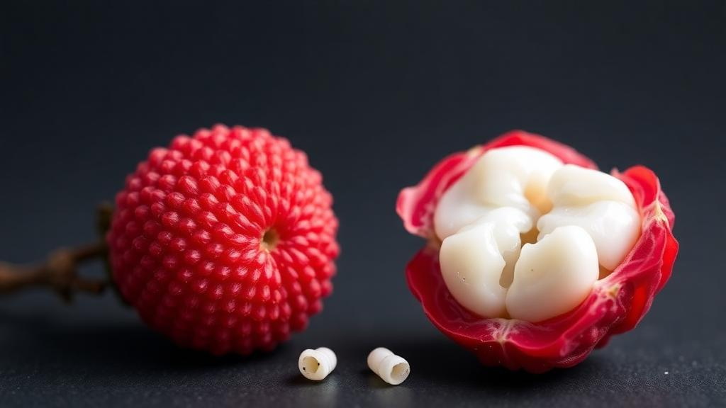
A discarded fruit pit may hold the key to faster healing inside the mouth. In a laboratory study published in BMC Complementary Medicine and Therapies, researchers from Mahidol University, the University of Phayao, Siam University and the Institute of Science Tokyo report that a crude extract prepared from lychee seeds can stimulate the proliferation and migration of human gingival fibroblasts, the connective tissue cells that orchestrate wound repair in the gums. The work, led by Titikan Ruensukon and Pornpoj Fuangtharnthip, is the first to examine how lychee seed extract affects oral wound healing, extending earlier observations that the same extract improved healing of skin wounds. The findings are early-stage and confined to cell cultures, but they point toward a possible natural ingredient for future treatments of painful oral soft tissue lesions such as recurrent aphthous ulcers, better known as canker sores.
The clinical problem the team set out to address is a familiar one. Ulcers and other lesions on the oral mucosa interfere with chewing and speech and can cause considerable pain, and their repair depends heavily on the behavior of human gingival fibroblasts. These cells migrate into the wound bed, proliferate, and lay down new extracellular matrix, including collagen, that closes the defect. Oxidative stress, driven by reactive oxygen species, damages fibroblasts and slows this sequence of events, delaying recovery. Because lychee seeds are rich in antioxidant phenolic and flavonoid compounds, and because previous work had suggested benefits for dermal wound healing, the researchers hypothesized that a crude lychee seed extract might protect or even stimulate gingival fibroblasts in ways that could accelerate oral tissue repair.
To prepare the test material, the team produced a crude extract by soaking lychee seed material in 95 percent ethanol, a standard approach for pulling out a broad spectrum of plant secondary metabolites. They then characterized the chemical composition of the extract using ultra-high-performance liquid chromatography coupled to quadrupole time-of-flight tandem mass spectrometry, a sensitive analytical technique that separates compounds and identifies them by their mass fragmentation patterns. The analysis revealed 45 compounds spanning six chemical classes, among them 15 phenolic and flavonoid species. This chemical inventory matters because phenolics and flavonoids are the compounds most often associated with antioxidant and wound-healing activity in plant extracts, and knowing what is present provides a starting point for later work with purified constituents.
The biological experiments used the HGF-1 human gingival fibroblast cell line, a commercially available established line, treated with crude lychee seed extract across a wide concentration range from 0.1 to 100 micrograms per milliliter. Cytotoxicity was assessed with the MTT assay, a colorimetric test in which metabolically active cells convert a tetrazolium compound into a colored product whose absorbance can be measured, following the ISO 10993-5 standard for evaluating the biological safety of medical devices. Cell morphology was examined by phase-contrast microscopy to check for visible signs of damage or distress. According to the ISO threshold, cell viability above 70 percent indicates a non-cytotoxic material, and the extract met that criterion at concentrations from 0.1 up to 50 micrograms per milliliter.
At the highest concentration tested, 100 micrograms per milliliter, viability fell marginally below the 70 percent threshold, although the reduction did not reach statistical significance compared with untreated controls. The 50 and 100 microgram per milliliter doses also behaved differently in the functional assays, suppressing fibroblast growth rather than supporting it. This concentration-dependent pattern, in which low doses help and high doses hinder, is common in studies of plant extracts and reflects the dual nature of many plant polyphenols, which can act as protective antioxidants at low exposure but become pro-oxidant or otherwise disruptive at higher levels. For any future therapeutic application, the study suggests the useful window sits well below the cytotoxic boundary.
Proliferation was tracked with the MTT assay over seven days of continuous exposure. At low concentrations between 1 and 20 micrograms per milliliter, the extract significantly enhanced HGF-1 proliferation on day five. By day seven, the lowest dose tested, 0.1 micrograms per milliliter, produced the highest proliferation of all, reaching statistical significance at p less than 0.001 compared with the control. The authors analyzed the data with one-way analysis of variance followed by Tukey’s honestly significant difference post-hoc test, with significance set at p less than 0.05. The result that the most dilute dose ultimately drove the strongest growth response hints that the active compounds are potent and that more is not necessarily better, a theme that recurred in the migration experiments.
Migration, the other half of the wound-repair equation, was evaluated with an in vitro scratch assay, a widely used technique in which a sterile pipette tip drags a straight gap through a confluent cell monolayer, mimicking a wound, and researchers then photograph the gap at intervals to measure how quickly cells close it. The team measured gap closure at 8, 16 and 24 hours after treatment. Concentrations from 0.1 to 20 micrograms per milliliter accelerated closure of the scratch at the eight-hour mark, and the middle-range doses of 5 and 10 micrograms per milliliter produced the greatest closure by 24 hours. In contrast, the 50 and 100 microgram per milliliter doses delayed gap closure, reinforcing the picture that moderate doses support the migratory behavior fibroblasts need to invade a wound while high doses impair it.
Putting the numbers together, the authors identify 0.1 to 10 micrograms per milliliter as the most promising concentration range, one in which the crude extract is clearly non-cytotoxic, stimulates fibroblast proliferation, and speeds wound closure in the scratch model. They are careful about the limits of interpretation. The study demonstrated an association between extract exposure and improved cellular outcomes but did not determine the underlying molecular mechanisms. The extract contains a complex mixture, and any of its 45 identified compounds, alone or in combination, could be responsible for the effects. Possible pathways that the authors discuss in the broader context include antioxidant defense signaling through the Nrf2 pathway and growth factor signaling, but these remain hypotheses for future experiments rather than demonstrated findings of this study.
The researchers also frame the work within the biology of oral wound healing. Fibroblast proliferation and migration are early and essential steps in granulation tissue formation, and fibroblasts respond to signals such as transforming growth factor beta 1 and fibroblast growth factor 2 while producing collagen and remodeling the matrix with matrix metalloproteinases. Oxidative stress in the wound environment, elevated reactive oxygen species and inflammatory cytokines such as interleukin 1 beta, interleukin 6 and tumor necrosis factor alpha can all impair these processes. An extract rich in antioxidant phenolics could, in principle, shift that balance, which is why the authors suggest that crude lychee seed extract and its purified constituents merit further investigation as agents for promoting oral soft tissue healing.
For now, the caveats are substantial. Everything reported here happened in a dish, using a single established cell line and a crude, chemically unstandardized extract, and the authors note that no specific funding body supported the research and that they declare no competing interests. The jump from a scratch assay to a mouthwash, gel or dressing that safely accelerates ulcer healing in patients requires animal studies, mechanism work, extract standardization and eventually clinical trials. Still, the study opens an unexpected door. Lychee seeds are an abundant agricultural byproduct, largely thrown away after the fruit is eaten, and if their phenolic cargo can be harnessed for oral tissue repair, a tropical fruit pit could someday find an unlikely second life in dental medicine. The next step, the authors suggest, is to isolate and test the individual purified compounds responsible for the proliferative and migratory effects they observed.
Subject of Research: Effects of crude lychee seed extract on proliferation and migration of human gingival fibroblasts in vitro
Article Title: In vitro assessment of cell proliferation and migration induced by crude lychee seed extract in human gingival fibroblast cells (HGF-1)
Article References: Ruensukon, T., Chuenjitkuntaworn, B., Tengrungsun, T., Pachimsawat, P., Tamura, Y., & Fuangtharnthip, P. (2026). In vitro assessment of cell proliferation and migration induced by crude lychee seed extract in human gingival fibroblast cells (HGF-1). BMC Complementary Medicine and Therapies. https://doi.org/10.1186/s12906-026-05579-w
Image Credits: AI Generated
DOI: 10.1186/s12906-026-05579-w
Keywords: lychee seed extract, human gingival fibroblasts, wound healing, cell proliferation, cell migration, oral ulcers, natural products, phenolic compounds, flavonoids, MTT assay, scratch assay, cytotoxicity
News Source: Alan Morgan. (October 4, 2026). Lychee Seed Extract Shows Promise for Healing Oral Wounds in Lab Study. Scienmag.