Pulmonary tuberculosis has long been described as a disease with a signature: a cavity carved into the upper lobes of the lung, surrounded by consolidation, in a patient with a productive cough. But in people whose immune systems have been devastated by HIV, that signature can vanish. A new cross-sectional study from southern Vietnam, published in BMC Infectious Diseases, provides one of the most granular real-world pictures yet of how tuberculosis actually looks, sounds and tests in adults with advanced HIV disease, and it confirms a clinical suspicion that has circulated for decades: as CD4 cell counts fall, tuberculosis stops looking like tuberculosis.
The study, conducted by Bao Linh Nguyen and Thanh Binh Ngo of the University of Medicine and Pharmacy at Ho Chi Minh City, enrolled adults with HIV and microbiologically confirmed pulmonary tuberculosis at a tertiary referral hospital for tuberculosis and lung diseases between December 2022 and September 2023. Rather than averaging findings across the cohort, the researchers deliberately stratified patients by CD4 count, the standard laboratory measure of immune destruction, and tracked how three separate axes of the disease shifted across those strata: the radiographic appearance of the lungs on chest X-ray, the microbiological yield of sputum tests, and the presence of additional pulmonary pathogens sharing the same airways.
The patients themselves tell much of the story. The median CD4 count in the cohort was a startling 40.5 cells per microliter of blood, a level at which the immune system is barely functional; healthy counts sit above 500. Most patients were not yet taking antiretroviral therapy, and many were learning of their HIV diagnosis for the first time when they arrived at the hospital. This is the face of late presentation, and it is common in settings where HIV stigma, fragmented testing services and limited access to care delay diagnosis until opportunistic infections force patients through the door. Vietnam’s HIV epidemic, concentrated in certain regions and populations, continues to feed such late presentations into its tuberculosis wards.
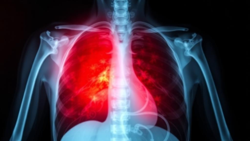
The radiographic findings were the study’s most striking result. Among patients in the highest CD4 stratum, every single patient showed cavitation on chest radiography, and nearly nine in ten showed consolidation, the dense opacities that mark the body’s attempt to wall off infection with an intact inflammatory response. Large nodules appeared in two-thirds of this group. In the lowest CD4 stratum, the picture inverted almost completely: cavitation was seen in just 3.8 percent of patients, consolidation in 30.8 percent, and large nodules in 9.6 percent. Instead, half of the most immunosuppressed patients displayed ground-glass opacity, a hazy, frosted-glass pattern that in this population strongly evokes Pneumocystis jirovecii pneumonia or other non-tuberculous pathology. All four of these radiographic trends remained statistically significant after correction for multiple comparisons, with adjusted q-values of 0.025 or lower.
The biological explanation is straightforward but consequential. Cavitation in tuberculosis is not the work of the bacterium alone; it is the work of the immune response to it. Caseous necrosis, the destruction of lung tissue into a cheese-like core that can liquefy and drain into airways, depends on a vigorous CD4 T-cell driven inflammatory reaction. When CD4 cells are scarce, the lung cannot mount that reaction, so the bacilli multiply diffusely without forming cavities, and the radiograph shows vague haziness rather than the textbook cavity. The practical implication is that clinicians who dismiss tuberculosis because the X-ray lacks classic features may be precisely wrong in the patients at greatest risk, since atypical, ground-glass-predominant disease is a marker of the deepest immunosuppression.
That overlap with Pneumocystis is where the study becomes clinically urgent. Ground-glass opacity showed an unadjusted association with Pneumocystis positivity, with an odds ratio of 5.67, meaning patients with this pattern were more than five times as likely to harbor the fungus. Yet the diagnostic workup for Pneumocystis was strikingly incomplete: among patients with CD4 counts at or above 200 cells per microliter, only four of nine were ever tested, and none of those tested was positive. Overall, at least one additional pulmonary organism was documented in 28 of 92 patients, or 30.4 percent of the cohort. In other words, nearly a third of these patients were fighting more than one lung infection simultaneously, a reality that complicates every treatment decision, from empiric antibiotic choices to the timing and composition of antiretroviral initiation, where immune reconstitution inflammatory syndrome looms as a hazard.
On the microbiological axis, the study delivered a clear verdict on diagnostics. Sputum smear microscopy, the century-old workhorse of tuberculosis diagnosis in high-burden countries, detected acid-fast bacilli in only 63.3 percent of patients. The molecular Xpert MTB/RIF assay, which amplifies Mycobacterium tuberculosis DNA and simultaneously screens for rifampicin resistance, was positive in 91.1 percent. The paired comparison, analyzed with exact McNemar testing, produced a p-value below 0.0001, leaving little doubt that the molecular assay outperforms smear across every level of immunosuppression. Notably, neither test showed a significant ordered trend across CD4 strata, suggesting that the well-known paucibacillary appearance of tuberculosis in advanced HIV does not translate into a simple, predictable gradient of test positivity in real-world sputum samples.
Perhaps the most pragmatic finding concerns a test that almost any district laboratory can perform. The researchers explored whether the absolute lymphocyte count, a routine component of a complete blood count, could serve as a proxy for CD4 depletion. In exploratory receiver-operating-characteristic analysis, the absolute lymphocyte count discriminated patients with CD4 counts below 200 cells per microliter with an area under the curve of 0.89, with a 95 percent confidence interval of 0.79 to 0.96. An AUC near 0.9 represents strong discriminatory power, approaching the threshold often considered excellent. The authors are careful to frame this correctly: the lymphocyte count may provide a supportive indicator of advanced immunosuppression where CD4 testing infrastructure is unavailable, but it must not substitute for direct CD4 measurement, which remains the gold standard for staging HIV disease and guiding prophylaxis against opportunistic infections.
The study’s limitations deserve honest weight, and the authors acknowledge them directly. This was a single-center, cross-sectional investigation at a specialized referral hospital, which means the patients may not represent the broader population of people with HIV and tuberculosis in Vietnam or elsewhere. The design captures a snapshot rather than a trajectory, so it can describe associations but cannot establish how individual patients’ disease evolves as immune function changes. The Pneumocystis findings rest on microscopy, an insensitive method, and the testing gaps among higher-CD4 patients make the true prevalence of coinfection uncertain. The lymphocyte count analysis was explicitly exploratory. The authors describe their conclusions as hypothesis-generating and call for prospective confirmation, a framing that reflects appropriate scientific caution rather than weakness.
Even with those caveats, the findings land at a consequential moment. Tuberculosis remains the leading cause of death among people with HIV worldwide, and the World Health Organization’s targets for ending the dual epidemic depend on finding cases earlier and treating them correctly the first time. This study from Ho Chi Minh City distills that challenge into concrete clinical rules of thumb: in a patient with advanced HIV, a hazy chest X-ray without cavitation should raise, not lower, suspicion of tuberculosis; molecular testing should replace smear wherever possible; a substantial fraction of patients harbor secondary pulmonary pathogens that demand broader diagnostic thinking; and where CD4 counters are scarce, a simple lymphocyte count can flag the patients most likely to be dangerously immunosuppressed. For the clinicians working on the front lines of the HIV-tuberculosis collision, these are not abstractions. They are the difference between a diagnosis made in time and one made too late.
Subject of Research: Clinical, radiographic and microbiological presentation of HIV-associated pulmonary tuberculosis across CD4 count strata
Article Title: Clinical, radiographic and microbiological manifestations of newly diagnosed pulmonary tuberculosis across CD4 strata in adults with advanced HIV disease: a real-world cross-sectional study in southern Vietnam
Article References: Nguyen, B. L., & Ngo, T. B. (2026). Clinical, radiographic and microbiological manifestations of newly diagnosed pulmonary tuberculosis across CD4 strata in adults with advanced HIV disease: a real-world cross-sectional study in southern Vietnam. BMC Infectious Diseases. https://doi.org/10.1186/s12879-026-14507-9
Image Credits: AI Generated
DOI: 10.1186/s12879-026-14507-9
Keywords: pulmonary tuberculosis, HIV, advanced HIV disease, CD4 count, chest radiography, ground-glass opacity, Xpert MTB/RIF, sputum smear microscopy, Pneumocystis jirovecii, coinfection, absolute lymphocyte count, Vietnam
News Source: Kristina Jarvis. (October 4, 2026). As Immunity Falls, Tuberculosis in HIV Wears a Different Face on the Chest X-Ray. Scienmag.