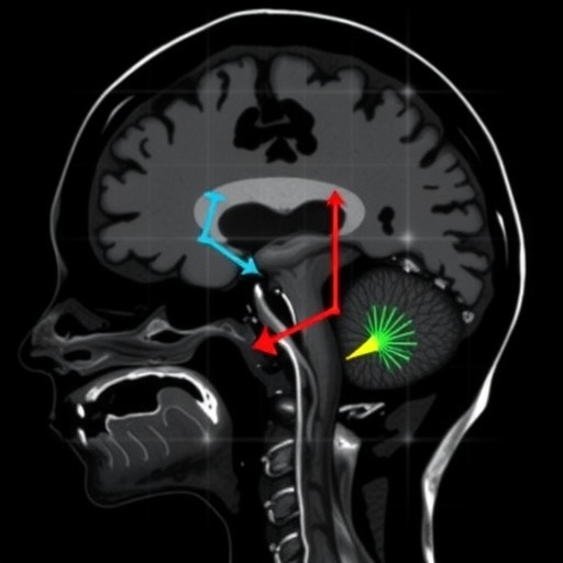
In a rare, comprehensive new guide published in Pediatric Radiology, a leading pediatric neuroradiologist has laid out the full spectrum of spinal cerebrospinal fluid (CSF) leaks in children—a condition so uncommon that it is often missed, sometimes for years, leaving young patients with debilitating headaches, hearing problems, and other neurological symptoms that baffles multiple specialists before anyone identifies the true cause. The review, authored by Mark D. Mamlouk of Kaiser Permanente Santa Clara Medical Center and the University of California, San Francisco, offers clinicians a practical, image-rich framework for recognizing, classifying, and treating these elusive leaks, and it arrives at a moment when the field’s understanding of CSF leak physiology is advancing faster than ever.
Spinal CSF leaks occur when the fluid that cushions the brain and spinal cord escapes through a defect in the dura, the tough membrane that encloses the central nervous system. The loss of fluid leads to intracranial hypotension—abnormally low pressure inside the skull. The hallmark symptom is an orthostatic headache, one that worsens when a child stands upright and eases when they lie down. Valsalva maneuvers such as coughing, sneezing, or bending over can intensify the pain. But the clinical picture is rarely so tidy. Not all children present with positional headaches, and the headaches can fade over time, causing clinicians to overlook the diagnosis entirely. Additional symptoms may include neck pain, muffled hearing, tinnitus, vertigo, and even cranial neuropathies. Because symptoms so often mimic more common conditions, children may spend months or years bouncing between specialists before the leak is identified.
The new review classifies pediatric spinal CSF leaks into two broad categories: nonspontaneous leaks, which follow trauma or medical procedures, and spontaneous leaks, which arise without obvious cause. Nonspontaneous leaks are frequently iatrogenic—the result of a lumbar puncture, an epidural catheter placement, or surgery. A needle puncture most commonly tears the dura dorsally, but if the needle passes all the way through the thecal sac, the resulting tear may be ventral, requiring a very different treatment approach. A lesser-known iatrogenic entity is the arachnoid bleb, in which a small pouch of arachnoid membrane herniates through a dural rent created by a needle. Traumatic leaks, meanwhile, can occur anywhere along the neuroaxis, including the sacrum, and typically appear on MRI as extradural fluid collections.
Spontaneous leaks in children fall into three distinct types: dural tears, CSF-venous fistulas, and CSF-vascular malformation fistulas. Dural tears may be ventral or lateral, and on spine MRI they produce extradural fluid that begins as unorganized collections in acute stages but can evolve into organized, membrane-encased collections in chronic stages as neo-membranes form around the leaked fluid. Notably, the review argues that extradural arachnoid cysts—long considered a separate entity—may actually represent a form of lateral dural tear, with recent research suggesting these lesions are better described on imaging as extradural collections rather than true cysts. One striking case in the review involves an eight-year-old boy with enuresis and constipation whose dorsal thoracolumbar collection remodeled the posterior elements of his vertebrae; digital subtraction myelography pinpointed a contrast jet at the left T12 nerve root, and targeted surgery to repair the dural defect cured him.
CSF-venous fistulas (CVFs) represent an abnormal communication between the subarachnoid space and a spinal vein, allowing CSF to drain directly into the venous system. These are increasingly recognized in adults—indeed, at least one adult study found CVFs more common than dural tears—but they appear to be exceedingly rare in children, where dural tears dominate. The third category, CSF-vascular malformation fistulas, involves unregulated shunting from the spine into a venous or lymphatic malformation, and the review documents a remarkable case in which a fistula first appeared in a sixteen-month-old girl who abruptly stopped walking—initially misdiagnosed as an epidural hematoma—and was only correctly identified twenty-two years later when she presented with headaches and a presumed “Chiari” malformation.
Connective tissue disorders are a significant risk factor in pediatric cases. Marfan syndrome, for instance, can produce dural fragility that predisposes children to spontaneous leaks, often accompanied by sacral dural ectasia visible on imaging. Children with diffuse or paraspinal vascular malformations face elevated risk of CSF-vascular malformation fistulas. The rarity of pediatric leaks overall—spontaneous intracranial hypotension occurs in at least 4 to 5 per 100,000 adults, and less frequently in children—means most clinical knowledge is extrapolated from adult populations, a gap the review aims to help close.
Diagnosis rests on a carefully sequenced imaging algorithm. Contrast brain MRI is the first-line study for suspected intracranial hypotension, with sagittal imaging assessing brain sag and venous distension, pre- and post-gadolinium T1-weighted sequences detecting dural enhancement, and T2 or FLAIR sequences evaluating subdural collections and alternative diagnoses. Optic nerve sheath fluid is another key indicator—when the sheath diameter falls below roughly 4.4 millimeters, an underlying CVF becomes more likely. The SIH Bern score aggregates these findings into a probability assessment: quantitative measurements of the suprasellar cistern (≤4 mm, 2 points), prepontine cistern (≤5 mm, 1 point), and mamillopontine distance (≤6.5 mm, 1 point), combined with qualitative features including dural enhancement (2 points), venous sinus distension (2 points), and subdural collections (1 point), categorize patients as low (0–2), intermediate (3–4), or high (≥5) probability for an underlying leak. Critically, the Bern score was validated only in adults, and the review cautions that its pediatric utility remains unproven.
On spine MRI, the pivotal finding is the spinal longitudinal extradural collection (SLEC)—T2-hyperintense fluid within the epidural space, often spanning multiple levels, best seen on fat-suppressed sequences where it displaces the normally dark epidural fat. The presence or absence of a SLEC determines the downstream pathway. If a SLEC is present, pointing to a dural tear, the patient undergoes CT myelography positioned prone in Trendelenburg for suspected ventral leaks, or in decubitus Trendelenburg for suspected lateral leaks, using small 1–2 milliliter boluses of preservative-free iohexol contrast. A ventral tear appears as a split contrast column extending from the subarachnoid to the ventral epidural space. If no SLEC is seen, decubitus myelography with 5–8 milliliters of contrast searches for CVFs along the entire spine, with the option of rotating the patient to the contralateral side for a bilateral assessment.
The review pays particular attention to pediatric-specific considerations. Ionizing radiation from myelography demands dose minimization—scanning should be concentrated over the SLEC levels rather than repeatedly imaging the whole spine as is sometimes done in adults. Digital subtraction myelography may offer lower radiation exposure than CT myelography while preserving diagnostic accuracy. Contrast dosing must respect age-specific iodine limits, and sedation or general anesthesia is often necessary, requiring careful coordination with anesthesia teams regarding prone and Trendelenburg positioning. A particularly instructive pitfall is the confusion between intracranial hypotension and Chiari 1 deformity: both can cause cerebellar tonsillar descent, and Chiari 1 can even develop during childhood as the skeleton grows, mimicking acquired brain sag. The mamillopontine distance is the decisive discriminator—reduced in intracranial hypotension, preserved in Chiari 1.
Treatment options span three approaches. Epidural blood patching, performed under CT or fluoroscopic guidance, delivers autologous blood to the dural defect; for ventral tears, needles are advanced through the lower third of the neural foramen to avoid the vasculature and nerves in the upper third. The review emphasizes that the traditional 20-milliliter blood volume, drawn from obstetric literature, should not be treated as a fixed requirement—patching volume should be individualized to leak location, patient size, and imaging appearance. Fibrin glue, though off-label, offers stronger adhesive sealing properties and can be used alone or with blood. For CVFs, targeted fibrin glue patching at the cyst-vein junction or transvenous embolization—selectively catheterizing the involved vein and injecting embolic material—are both viable, though no direct comparisons exist. Surgery remains the definitive option for any leak type, particularly ventral dural tears with organized collections and arachnoid blebs that resist patching.
Perhaps the review’s most clinically valuable contribution is its guidance on patients with negative MRI findings. In children, unlike adults, most true CSF leaks will show abnormalities on brain MRI, spine MRI, or both; definitive CVFs with entirely normal imaging are the exception. When initial MRIs are normal and suspicion is low to intermediate, the review advises careful re-review of subtle findings like venous distension before considering myelography or empiric patching—and stresses that unnecessary invasive procedures in children should be minimized. With modern myelography, image-guided patching, and endovascular therapies now mature, the neuroradiologist has moved from a purely diagnostic role to the center of both detection and cure for this hidden epidemic of childhood headache.
Subject of Research: Pediatric spinal cerebrospinal fluid (CSF) leaks, including their classification, imaging diagnosis, and treatment
Subject of Research: Cancer
Article Title: Detecting the drip: pediatric neuroradiologist guide to spinal CSF leaks
Article References: Mamlouk, M. D. (2026). Detecting the drip: pediatric neuroradiologist guide to spinal CSF leaks. Pediatric Radiology. https://doi.org/10.1007/s00247-026-06764-2
Image Credits: AI Generated
DOI: 10.1007/s00247-026-06764-2
Keywords: Pediatric spinal CSF leaks, Spontaneous intracranial hypotension, CSF-venous fistula, Dural tear, Epidural blood patch, Fibrin glue patching, CT myelography, Digital subtraction myelography, Brain MRI, Bern score, Chiari 1 deformity, Neuroradiology
Cite Scienmag News
APA MLA Chicago
Nathaniel Bowman. (September 6, 2026). Spotting spinal CSF leaks in children: a neuroradiologist’s guide. Scienmag. https://scienmag.com/spotting-spinal-csf-leaks-in-children-a-neuroradiologists-guide/
Nathaniel Bowman. “Spotting spinal CSF leaks in children: a neuroradiologist’s guide.” Scienmag, 6 September 2026, https://scienmag.com/spotting-spinal-csf-leaks-in-children-a-neuroradiologists-guide/. Accessed 6 September 2026.
Nathaniel Bowman. “Spotting spinal CSF leaks in children: a neuroradiologist’s guide.” Scienmag. September 6, 2026. https://scienmag.com/spotting-spinal-csf-leaks-in-children-a-neuroradiologists-guide/
Copy citation Download RIS
Tags: advanced imaging in pediatric neurodiagnosticscerebrospinal fluid leak diagnosiscomprehensive guide to pediatric spinal leaksimaging techniques for CSF leaksMRI imaging for CSF leaksneuroimaging frameworks for CSF leaksneurological symptoms in childrenneuroradiologist guide to CSF leaksneuroradiology in childrenorthostatic headache in pediatricspediatric headache diagnosispediatric intracranial hypotensionpediatric neuroradiologypediatric spinal CSF leak diagnosispediatric spinal CSF leaksrare causes of pediatric headacherare neurological conditions in childrenspinal cerebrospinal fluid leak detectionspinal dural defects in childrentreatment approaches for pediatric CSF leakstreatment of pediatric CSF leaksValsalva maneuver effects on CSF leaks