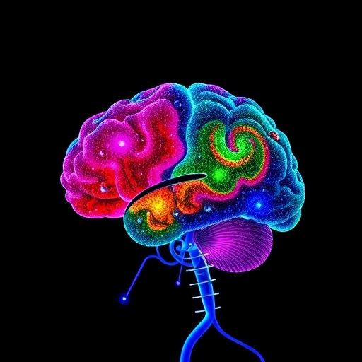
A new artificial intelligence model could help pathologists identify the molecular subtypes of central nervous system tumors using information hidden in ordinary microscope images, according to a study published in Nature Cancer. The model, named Hetairos, is designed to predict tumor methylation subtypes from histology—the study of tissue structure—rather than relying exclusively on the specialized molecular tests that currently define many brain tumor diagnoses. The work by D. Jin, A. Shmatko, A. Patel and colleagues points toward a future in which a scanned pathology slide may provide an early, highly informative guide to the biological identity of a tumor.
Central nervous system tumors are not defined solely by what they look like under a microscope. Two tumors that appear similar in tissue sections can behave very differently because their genomes, epigenomes and cellular programs differ. Modern classifications therefore incorporate molecular features, including patterns of DNA methylation. In this context, methylation refers to chemical tags attached to DNA that influence how genes are regulated without changing the underlying genetic sequence. Across tumor cells, these tags form recognizable patterns that can act like molecular fingerprints, helping distinguish clinically meaningful disease subtypes.
Methylation-based classification has become particularly important in neuro-oncology because the central nervous system contains a wide variety of tumors with overlapping appearances but sharply different prognoses and treatment strategies. A conventional histological diagnosis can remain uncertain when a sample is small, damaged, unusually differentiated or taken from a tumor with an uncommon molecular profile. Laboratory methylation profiling can resolve some of these cases, but it requires dedicated assays, specialized computational analysis and additional time. Hetairos addresses this gap by attempting to infer those molecular categories from the visual architecture preserved in stained tissue.
The model’s central premise is that molecular identity leaves visible traces. Tumor cells with different genetic and epigenetic programs may grow in different arrangements, alter the surrounding tissue in distinctive ways, form characteristic blood vessels or produce recognizable patterns of necrosis and cellular density. These features may be subtle, distributed across a slide and difficult for even experienced observers to weigh consistently. An artificial intelligence system can examine thousands or millions of image regions, measure relationships among cells and structures, and combine these signals into a prediction that is difficult to reproduce through unaided visual inspection.
A histology-based model typically begins with a digitized slide, created by scanning a glass tissue section at high resolution. The image is divided into smaller regions so that a neural network can learn local features such as nuclear shape, texture and cell density, while also recognizing broader patterns across the tumor. During training, the system is shown examples linked to reference methylation classifications. It gradually adjusts millions of internal parameters to associate visual patterns with molecular labels. Once trained, the model can generate a probability distribution across possible subtypes rather than simply issuing a single unexplained answer.
That distinction is important clinically. A prediction is most useful when it communicates confidence and identifies cases that require additional testing. If a slide contains features associated with several classes, or if the tissue differs substantially from the examples used during training, a responsible system should signal uncertainty rather than present a potentially misleading definitive diagnosis. The practical value of Hetairos will therefore depend not only on whether it can make accurate predictions, but also on how reliably it recognizes unfamiliar tumors, poor-quality samples and cases outside its training distribution.
The approach could eventually streamline the diagnostic pathway. Histological slides are already produced as part of routine pathology, meaning that an image-based model may be able to provide information without consuming another tissue section or waiting for a separate molecular assay. A rapid prediction could help prioritize confirmatory testing, alert clinicians to a tumor requiring urgent molecular characterization and support review of rare or ambiguous cases. In hospitals with limited access to advanced methylation laboratories, such software might also broaden access to molecularly informed diagnosis, provided it is rigorously validated and integrated with expert oversight.
Yet the model does not eliminate the need for molecular testing or pathologists. An image can contain clues to methylation status, but it is not itself a direct measurement of DNA methylation. Tissue preparation, staining protocols, scanner settings, magnification and differences among hospitals can all affect the appearance of a slide. Artificial intelligence systems may also learn accidental correlations, such as laboratory-specific artifacts or demographic patterns, instead of the underlying biology. A model that performs strongly on data from one institution may lose accuracy when applied to slides prepared elsewhere. Independent testing across hospitals, scanners, populations and tumor types is therefore essential before clinical deployment.
The broader significance of Hetairos lies in its attempt to connect two traditionally separate layers of cancer diagnosis: morphology and molecular biology. Pathology images preserve the spatial organization of disease, while methylation profiles capture regulatory states that are invisible to ordinary microscopy. By linking the two, AI could transform the microscope slide from a descriptive record into a computationally interpretable molecular proxy. That possibility is especially striking in brain tumors, where obtaining tissue can be difficult and where precise classification can influence surgery, radiation, chemotherapy, surveillance and discussions about prognosis.
The study arrives as computational pathology moves from experimental demonstrations toward tools intended to support real clinical decisions. Its success will ultimately be measured not by viral attention or visually impressive predictions, but by reproducibility, transparency and patient benefit. The authors’ description of Hetairos presents a model built to predict central nervous system tumor methylation subtypes from histology, an ambitious step toward faster molecular triage. If future studies confirm that its predictions remain reliable in diverse real-world settings, a routine tissue image could become an unexpectedly powerful first signal of a tumor’s hidden molecular identity.
Subject of Research: Histology-based artificial intelligence prediction of central nervous system tumor methylation subtypes
Article Title: Hetairos is a histology-based artificial intelligence model for predicting central nervous system tumor methylation subtypes
Article References: Jin, D., Shmatko, A., Patel, A. et al. Hetairos is a histology-based artificial intelligence model for predicting central nervous system tumor methylation subtypes. Nature Cancer 7, 884–898 (2026). https://doi.org/10.1038/s43018-026-01186-3
Image Credits: AI Generated
DOI: https://doi.org/10.1038/s43018-026-01186-3
Keywords: artificial intelligence, digital pathology, histology, central nervous system tumors, brain tumors, DNA methylation, tumor classification, computational pathology, neuro-oncology
Tags: AI model for tumor methylation subtype predictionbrain tumor classificationcomputer-aided pathology for tumor classificationdigital pathology and molecular diagnosticsDNA methylation patterns in brain tumorsearly detection of CNS tumor subtypes using AIHetairos artificial intelligence in neuro-oncologyhistology-based tumor diagnosisintegrating histology and methylation data in neuro-oncologymachine learning for brain tumor subtypesmolecular subtyping of central nervous system tumorstumor epigenome analysis using AI