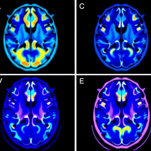
A new study reports that functional MRI can reveal how deep brain stimulation (DBS) in the globus pallidus internus (GPi) engages distinct, target-dependent brain circuits. The work, led by Santyr and colleagues, uses active GPi DBS while participants undergo fMRI, offering a systems-level view of stimulation effects that goes beyond electrode placement and clinical symptom reports.
The researchers focus on the idea that GPi is not a single “switch” for movement, but a hub whose activation patterns depend on where and how stimulation is delivered. By coupling real-time experimental stimulation states with imaging, the team sought to map network changes that emerge when DBS is engaged.
Technically, participants experienced stimulation during scanning, allowing the investigators to compare brain activity patterns under active versus non-active conditions. This design helps isolate stimulation-evoked responses, reducing the ambiguity that often arises when DBS is studied only through post hoc clinical outcomes or animal electrophysiology.
The key finding is that active GPi DBS produces network signatures that vary with the stimulation target. Instead of activating one uniform pathway, the imaging data show that distinct stimulation sites recruit different combinations of cortical and subcortical regions. Such target-specific network engagement may help explain why patients can benefit differently depending on parameter selection and anatomical targeting.
The study also emphasizes that DBS effects are distributed. Regions implicated in motor control and cognitive processing appear in the stimulation-evoked patterns, suggesting that GPi modulation can propagate through functional networks rather than acting only locally at the electrode site.
By identifying these network differences, the results provide a potential route toward more individualized DBS programming. If fMRI can reliably distinguish target-specific circuit engagement, clinicians may one day use imaging biomarkers to refine stimulation placement and settings for each patient.
Importantly, the approach moves the field toward “network-aware” neuromodulation. Rather than treating GPi as a static target, the findings argue for viewing DBS as an intervention that reshapes connectivity in a site-dependent manner.
Overall, the work frames viral science news worthy of immediate attention: mapping active DBS effects with fMRI could accelerate the translation of circuit-based optimization, improving both efficacy and mechanistic understanding for Parkinson’s disease therapies. The study appears in npj Parkinsons Diseases in 2026 (doi: 10.1038/s41531-026-01476-8).
Subject of Research: Functional MRI mapping of active GPi deep brain stimulation and target-specific brain networks in Parkinson’s disease
Article Title: fMRI during active GPi DBS uncovers target-specific networks
Article References: Santyr, B., Chow, C., Germann, J. et al. npj Parkinsons Dis. (2026). https://doi.org/10.1038/s41531-026-01476-8
Image Credits: AI Generated
Tags: brain network mapping during stimulationcortical and subcortical engagement in DBSfMRI in deep brain stimulationfunctional MRI for neuromodulationglobus pallidus internus neural circuitsmovement disorder neural pathwaysnetwork signatures of deep brain stimulationpersonalized neuromodulation mechanismsreal-time brain imaging during DBSstimulation site-dependent brain activationsystems-level analysis of DBS effectstarget-specific brain network activation