In the rapidly evolving landscape of biomedical research, one of the most transformative advancements in recent years has been the integration of deep learning technologies in pathology. The latest groundbreaking study, published in Nature Communications by Neidlinger et al., introduces a sophisticated deep learning framework designed to revolutionize pathology image analysis, promising to significantly enhance diagnostic accuracy and workflow efficiency. This development is poised to alter the fundamental ways in which pathology laboratories operate worldwide, blending artificial intelligence with conventional histopathological methodologies.
Pathology, the cornerstone of diagnostic medicine, relies heavily on the meticulous examination of tissue samples. Traditionally, this process demands extensive expertise and is often time-consuming, constrained by the subjective interpretation of pathologists. The study by Neidlinger and colleagues addresses these limitations head-on by harnessing deep learning, a subset of machine learning emphasizing neural networks capable of learning from large amounts of data. Their framework automates the complex task of analyzing high-resolution pathology images, enabling rapid and reliable interpretation of histopathological features that might otherwise be challenging to discern.
At the core of this innovative framework is an architecture optimized for handling the extraordinary scale and detail captured in pathology whole slide images. These images can encompass gigapixels of data, with intricate cellular and tissue structures that embody critical diagnostic information. The authors have engineered a neural network paradigm that not only copes with this vast data load but also excels in identifying morphological patterns indicative of various pathologies. The system’s design cleverly integrates multi-scale feature extraction, allowing it to understand cellular environments both in isolation and as part of the broader tissue context.
A key technical achievement of the study lies in the model’s ability to learn from relatively small datasets without compromising performance—a notorious challenge in medical image analysis due to the often-limited availability of labeled data. By incorporating advanced transfer learning techniques and data augmentation strategies, the framework generalizes effectively across different diseases and tissue types. This adaptability is particularly vital in pathology, where inter-patient heterogeneity and staining variations frequently blur the diagnostic picture.
The rigorous validation of the deep learning framework involved an impressively diverse set of pathology specimens, encompassing a range of cancers and inflammatory conditions. The authors demonstrate that their model outperforms traditional image analysis algorithms and even matches or exceeds the diagnostic accuracy of expert pathologists in several key tasks. These results underscore the potential for AI-driven pathology tools to act not just as assistants but as equal partners in clinical decision-making, expanding capabilities while reducing human error.
Importantly, the framework is engineered for seamless integration into existing digital pathology workflows. It supports interoperability with standard slide scanning hardware and software platforms, facilitating its adoption without demanding substantial infrastructural changes. This practical focus addresses a major hurdle in the clinical translation of AI technologies, which often falter due to integration challenges and workflow disruptions.
Another compelling aspect of the study is the system’s interpretability features. Unlike many black-box AI models, the framework provides visual explanations of its diagnostic decisions, highlighting the image regions most influential to its predictions. This transparency builds trust among clinicians and provides valuable insights for further validation and refinement of the model. Such interpretable AI is critical for meeting regulatory standards and encouraging widespread clinical acceptance.
The implications of this research extend well beyond the pathology department. The implementation of this deep learning framework can accelerate drug development, where precise tumor characterization is essential for patient stratification and treatment efficacy evaluation. Additionally, it opens avenues for telepathology, where digital slides analyzed by AI can support remote diagnosis in underserved areas, bridging critical gaps in healthcare access.
Neidlinger and colleagues also emphasize the scalability of their framework, an essential feature for adapting to future increases in digital pathology data volume driven by population growth and expanding screening programs. The computational efficiency of the model, achieved through algorithmic optimizations, ensures that the framework remains practical even in high-throughput clinical settings, preventing bottlenecks that could hinder patient care.
The intersection of deep learning with pathology image analysis exemplifies a vanguard approach in precision medicine, where computational tools augment human expertise to deliver personalized and timely diagnoses. As AI technologies mature and integrate further with clinical practice, they promise to elevate the standards of medical accuracy and efficiency, ultimately translating into improved patient outcomes and reduced healthcare costs.
Despite these advances, the authors acknowledge ongoing challenges, including the need for standardization in data preprocessing and annotation, which remain pivotal for training robust models. Moreover, ethical considerations surrounding AI in clinical decision-making, data privacy, and the medicolegal implications of machine-derived diagnoses form critical frontiers that parallel technological progress.
Looking ahead, the research community anticipates that frameworks like the one introduced by Neidlinger et al. will catalyze the development of even more sophisticated multi-modal AI systems, capable of integrating pathology images with genomic and clinical data. Such convergence promises to unveil deeper insights into disease mechanisms and facilitate truly personalized treatment strategies that combine histological, molecular, and patient-level information.
This study represents a decisive step toward the goal of democratizing access to advanced diagnostic capabilities through AI. By offering a highly efficient, interpretable, and adaptable deep learning framework tailored for pathology image analysis, Neidlinger and collaborators have opened new horizons in digital pathology. As this technology transitions from research environments to clinical routine, its potential to transform patient care and biomedical research is profound.
In sum, this pioneering work not only showcases the immense power of deep learning in tackling historically challenging problems in pathology but also sets a new benchmark for future AI applications in medicine. With continued refinement, validation, and ethical governance, such innovations will undoubtedly become indispensable tools in the armamentarium of modern healthcare.
Subject of Research: Deep learning applications for pathology image analysis in medical diagnostics.
Article Title: A deep learning framework for efficient pathology image analysis.
Article References:
Neidlinger, P., Lenz, T., Foersch, S. et al. A deep learning framework for efficient pathology image analysis. Nat Commun 17, 5740 (2026). https://doi.org/10.1038/s41467-026-74918-9
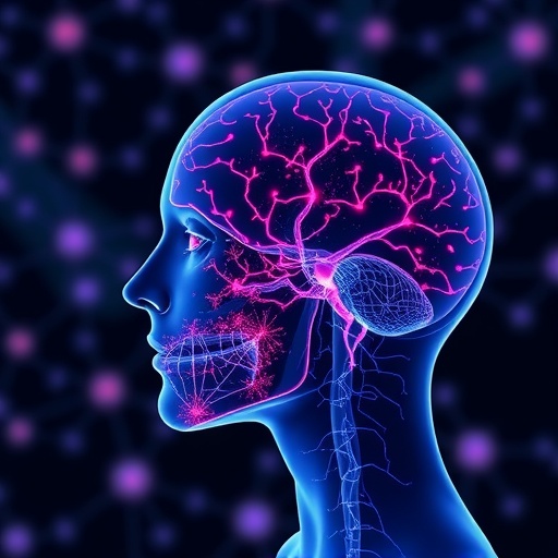
Image Credits: AI Generated
DOI: https://doi.org/10.1038/s41467-026-74918-9
Tags: AI in histopathologyartificial intelligence in tissue sample examinationautomated histopathological feature detectiondeep learning framework for pathology image analysisdeep learning in pathologyenhancing diagnostic accuracy with AImachine learning in diagnostic medicineneural networks for biomedical image analysispathology image automationpathology workflow efficiency improvementscalable deep learning architectures for pathologywhole-slide image analysis
