In a groundbreaking advance that promises to reshape the landscape of biomedical imaging, Zhou, Yang, He, and colleagues have unveiled a novel class of red-light-excited dynamic near-infrared (NIR) organic afterglow materials engineered specifically for in vivo bioimaging applications. Published in Light: Science & Applications, this 2026 study marks a significant leap forward in organic afterglow technology—materials that emit light for extended periods after excitation—opening new horizons for non-invasive, high-resolution imaging deep within living organisms.
The development of these materials addresses a longstanding challenge in biomedical optics: achieving deep tissue imaging with minimal phototoxicity and background noise. Traditional fluorescence imaging often relies on ultraviolet or blue light excitation, which suffers from limited tissue penetration and can induce cellular damage. By contrast, red and near-infrared light sources offer greater penetration and lower phototoxicity, but engineering organic materials that respond efficiently to such excitation while providing sustained afterglow has proven elusive—until now.
Central to the study is the creation of organic afterglow materials that are dynamically excitable by red light, enabling persistent near-infrared emission. This long-lived luminescence after cessation of excitation circumvents real-time excitation challenges, substantially reducing autofluorescence and scattering problems commonly faced in live tissues. The materials’ organic nature also circumvents potential biocompatibility concerns associated with heavy-metal-based inorganic phosphors, making them particularly suited for in vivo applications.
The researchers employed an innovative molecular design strategy that integrates specific chromophores with optimized energy states to facilitate efficient triplet harvesting and intersystem crossing. By finely tuning molecular structures, they achieved an organic afterglow system capable of robust red-light excitation, which then produces prolonged NIR emission. This dual-wavelength functionality is crucial for penetrating biological tissues and capturing high-contrast images over extended periods.
Detailed photophysical analyses revealed that these materials exhibit impressive afterglow lifetimes spanning seconds to minutes, maintaining emission intensity well beyond the duration of excitation. Such extended afterglow behavior is instrumental for practical imaging since it allows temporal separation between excitation and signal acquisition, thereby eliminating background fluorescence and enhancing signal-to-noise ratios.
Beyond their remarkable luminescence characteristics, these organic afterglow materials demonstrate excellent biocompatibility and stability in physiological environments. The team conducted extensive in vivo experiments to evaluate bioimaging performance, leveraging small animal models to visualize biological structures with unprecedented clarity and depth. The materials’ dynamic excitation capability permitted selective imaging of tissues without continuous light exposure, mitigating heat generation and photodamage risks.
One of the striking demonstrations involved tracking tumor tissues in live animals, where the afterglow probes illuminated cancerous regions with high spatial resolution and low invasiveness. This breakthrough suggests a powerful tool for early cancer detection and monitoring therapeutic responses, leveraging the materials’ capacity for deep tissue visualization without reliance on external dyes or radioactive tracers.
Moreover, the study highlights the tunability of these afterglow systems, suggesting future avenues for custom designing materials tailored for specific wavelengths or targeting capabilities. This versatility could pave the way for multiplexed imaging platforms, combining multiple organic afterglow probes to monitor diverse biological processes simultaneously with minimal crosstalk.
The energy-efficient nature of the red-light excitation mechanism also holds great promise for portable and wearable biosensors. Unlike conventional fluorescence methodologies demanding bulky and energy-intensive excitation sources, these materials require only low-intensity red light to trigger sustained NIR emission, potentially enabling compact, battery-powered imaging devices for point-of-care diagnostics.
The researchers also emphasize the implications for longitudinal biological studies. Because the afterglow emission persists after excitation has ended, repetitive imaging sessions can be conducted with minimal disturbance to the subject, enhancing the feasibility of real-time monitoring of dynamic physiological changes or disease progression in live organisms.
On the technical front, the molecular architecture involves carefully balancing intersystem crossing efficiency with triplet state stabilization, ensuring prolonged phosphorescence without compromising emission brightness. Advanced spectroscopic techniques confirmed the materials’ unique dynamic excitation behavior, elucidating the underlying photophysical mechanisms that differentiate them from conventional fluorescent or phosphorescent probes.
From a materials science perspective, the synthesis protocols prioritize scalability and cost-effectiveness, employing organic components readily available and amenable to modification. This practical approach is expected to accelerate translation from laboratory research to commercial biomedical imaging applications.
The study’s coherent integration of chemistry, photophysics, and biomedical engineering sets a benchmark for future research aiming to harness organic afterglow materials for clinical and research-oriented bioimaging. The ability to safely and effectively image deep tissues through red-light activation and NIR emission promises to enhance diagnostic accuracy, therapeutic monitoring, and even guided surgery procedures.
Furthermore, ethical and safety assessments conducted alongside the imaging trials confirmed negligible cytotoxic effects and minimal immune responses, underscoring the suitability of these organic afterglow materials for repeated use in living subjects.
In conclusion, the work of Zhou and colleagues significantly broadens the toolkit for non-invasive biological imaging by combining the advantages of red-light excitation and dynamic NIR afterglow emission within organic molecular frameworks. Their findings are poised to inspire a new wave of innovations in biosensing, enabling clinicians and researchers to peer deeper into living systems with unprecedented clarity, safety, and temporal flexibility.
This breakthrough not only advances fundamental knowledge in organic luminescent materials but also underscores the critical role of interdisciplinary collaborations in addressing complex biomedical challenges. As ongoing research refines these materials and integrates them into multifunctional imaging platforms, the future of bioimaging promises to be brighter, deeper, and more insightful than ever before.
Subject of Research: Development of red-light-excited dynamic near-infrared organic afterglow materials for in vivo bioimaging.
Article Title: Red-light-excited dynamic near-infrared organic afterglow materials for in vivo bioimaging.
Article References:
Zhou, L., Yang, J., He, Z. et al. Red-light-excited dynamic near-infrared organic afterglow materials for in vivo bioimaging. Light Sci Appl 15, 271 (2026). https://doi.org/10.1038/s41377-026-02340-3
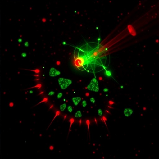
Image Credits: AI Generated
DOI: 10 June 2026
Tags: advanced organic afterglow technologybiomedical optics innovationdynamic near-infrared luminescencehigh-resolution biological imagingin vivo deep tissue imaginglow phototoxicity imaging agentsnon-invasive biomedical imaging techniquesorganic afterglow materials for bioimagingorganic materials for NIR emissionred and near-infrared excitation in bioimagingred-light-activated near-infrared afterglowreduced autofluorescence imaging


