In a groundbreaking revelation that challenges prevailing diagnostic paradigms in oncology, researchers from the University of Louisville School of Medicine have documented a remarkably rare and atypical manifestation of esophageal adenocarcinoma. Published in the March 2026 issue of Oncoscience, this pioneering case elucidates an esophageal tumor exhibiting exclusively extraluminal growth, invading the thoracic spine without any mucosal disruption detectable by conventional endoscopy. This extraordinary report not only expands our conceptual understanding of esophageal cancer presentation but also underscores critical pitfalls in current diagnostic approaches.
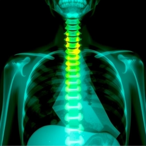
The patient in question, a 68-year-old male, initially presented with acute neurological deficits attributable to spinal cord compression at the T5 vertebra. This symptomatology included rapid onset lower-extremity weakness, sensory impairment, and urinary retention, indicating severe involvement of the central nervous system. Notably, these alarming neurologic signs emerged following several months of progressive dysphagia and significant weight loss, classical clinical harbingers of esophageal malignancy, yet paradoxically coupled with an unrevealing upper endoscopic evaluation.
Preceding hospital admission, the patient underwent thorough esophagogastroduodenoscopy complemented by extensive mucosal biopsies aimed at elucidating the cause of his dysphagia and cachexia. Surprisingly, the endoscopy revealed only benign findings related to reflux esophagitis and a hiatal hernia, with the esophageal and gastric mucosa grossly normal and devoid of malignancy or Barrett’s esophagus. This underscores the limitations of mucosal visualization in detecting submucosal or extraluminal tumor growth, particularly when the neoplasm bypasses the esophageal lumen entirely.
Subsequent advanced imaging dramatically altered the clinical landscape. Contrast-enhanced computed tomography delineated a large posterior mediastinal mass contiguous with the esophagus that had catastrophically eroded the T5 vertebral body, extending into the spinal canal and causing significant spinal cord compression. The imaging further revealed pulmonary nodules and hilar lymphadenopathy, suspicious for metastatic dissemination. This radiographic constellation vividly illustrated a tumor that, while silent endoscopically, was aggressively invading posterior anatomical structures typically spared in standard esophageal cancer progression.
Histopathological examination of the epidural tumor specimens obtained during urgent neurosurgical decompression confirmed the diagnosis of poorly differentiated adenocarcinoma consistent with an esophageal primary. Immunohistochemical profiling revealed positivity for cytokeratins AE1/AE3 and CK7, with patchy expression of CK20 and CDX-2, biomarkers strongly supportive of upper gastrointestinal tract origin. Importantly, pulmonary adenocarcinoma markers such as TTF-1 and Napsin A were negative, excluding a lung primary tumor. This detailed molecular fingerprint further corroborated the exceptional nature of tumor spread in this case.
This report delineates a previously uncharacterized pattern of esophageal adenocarcinoma—growth exclusive to the extraluminal space with direct invasion of posterior vertebral bodies. Traditionally, advanced esophageal cancers extend toward anterior or lateral mediastinal structures including the trachea, bronchi, aorta, or pleura, rarely exhibiting isolated posterior invasion. The tumor’s selective vertebral involvement, absent luminal mucosal changes detectable by endoscopy, is a novel finding that calls for a reevaluation of diagnostic strategies utilized in esophageal cancer.
The clinical implications of this case are profound. The reliance on endoscopic mucosal assessment alone could fail to detect submucosal or entirely extraluminal malignancies, particularly when patients persist with significant symptoms suggestive of esophageal cancer despite non-diagnostic initial investigations. This necessitates a high index of clinical suspicion accompanied by the incorporation of cross-sectional imaging modalities such as computed tomography and/or endoscopic ultrasound in persistent or equivocal cases to avoid diagnostic delays.
Furthermore, the case underscores the critical need for multidisciplinary collaboration among gastroenterologists, radiologists, oncologists, and neurosurgeons to address complex presentations of esophageal carcinoma. The urgent neurosurgical intervention undertaken — involving decompression and spinal stabilization — was instrumental in managing spinal cord compression and preventing further neurological deterioration, highlighting the importance of prompt intervention in such rare scenarios.
This singular presentation also accentuates the expansive heterogeneity of tumor biology within esophageal adenocarcinoma and suggests that neoplastic cells can adopt unconventional invasive trajectories, eluding traditional detection techniques. It challenges researchers and clinicians to investigate the molecular drivers and microenvironmental factors facilitating such rare patterns of tumor dissemination and invasion.
Moreover, this extraordinary case advocates for refinements in esophageal cancer staging and surveillance protocols, especially in high-risk individuals demonstrating persistent dysphagia and systemic symptoms with initial negative endoscopy. Integrating advanced diagnostic techniques can optimize early detection, improve prognostication, and guide timely therapeutic decisions.
By documenting this unprecedented extraluminal esophageal adenocarcinoma with posterior spinal invasion, the University of Louisville team has markedly extended the clinical spectrum of esophageal cancer. This knowledge compels the medical community to remain vigilant for atypical presentations, to adapt diagnostic workflows accordingly, and ultimately enhance patient outcomes through earlier intervention.
In conclusion, the remarkable documentation of an esophageal adenocarcinoma growing exclusively outside the lumen and invading the vertebral column despite a previously normal endoscopic examination demands paradigm shifts in clinical suspicion, diagnostic approaches, and collaborative care models. It is a clarion call to oncologists and gastroenterologists to broaden their evaluative lens when confronted with unresolved high-risk symptoms that defy conventional findings.
DOI: https://doi.org/10.18632/oncoscience.653
Correspondence: Benjamin Wenyuan Xie – [email protected]
Subject of Research: People
Article Title: Unprecedented non-luminal esophageal adenocarcinoma invading the spine
News Publication Date: May 14, 2026
Web References: https://doi.org/10.18632/oncoscience.653 ; https://www.oncoscience.us/archive/v13/
Image Credits: Copyright: © 2026 Xie and Kalantri. Distributed under the terms of CC BY 4.0 license.
Keywords: cancer, esophageal adenocarcinoma, atypical esophageal cancer, spinal invasion, non-luminal presentation, normal esophagogastroduodenoscopy
Tags: atypical esophageal tumor presentationchallenges in esophageal cancer diagnosisesophageal adenocarcinoma invading spineesophageal cancer and dysphagiaesophageal cancer with normal endoscopyesophageal cancer without mucosal disruptionextraluminal esophageal cancerlimitations of endoscopic biopsy in cancer detectionneurological deficits in esophageal cancerrare esophageal tumor growth patternsspinal cord compression symptomsthoracic spine tumor compression
