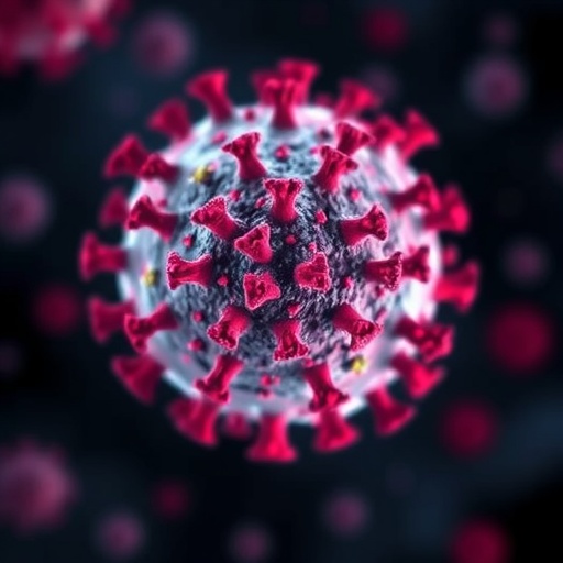
Emerging research from Tulane University has unveiled critical insights into the prolonged physiological consequences following mild infections of COVID-19 and influenza, deepening our understanding of why some patients endure lasting symptoms well beyond the acute phase of illness. This novel investigation elucidates differential tissue responses post-infection, shining light on the unique neuropathological aftermath attributable to SARS-CoV-2 in contrast to influenza virus.
The comprehensive study employed a murine model to dissect subchronic effects of both viral pathogens on pulmonary and cerebral tissues after the resolution of detectable viral load. Researchers meticulously characterized post-infectious landscapes, focusing on immune activation, fibrotic processes, and neuroinflammatory markers. The data revealed substantial parallels in lung injury mechanisms but striking disparities in brain pathology between the two infections.
Pulmonary analysis demonstrated that both SARS-CoV-2 and influenza instill a persistent state of immune cell activation, accompanied by collagen accumulation within lung parenchyma. Such fibrotic remodeling is known to compromise lung compliance and gas exchange efficiency, providing a plausible biological underpinning for prolonged dyspnea frequently observed clinically. Intriguingly, however, the lung’s intrinsic reparative response diverged markedly depending on the infective agent.
After influenza infection, researchers identified a robust activation of epithelial progenitor cells tasked with regenerating the airway lining, orchestrating tissue restitution and functional recovery. In stark contrast, this reparative cellular mobilization was conspicuously absent in SARS-CoV-2 infected lungs, implicating viral interference with regenerative pathways. This impairment may exacerbate long-term respiratory sequelae unique to COVID-19 convalescence.
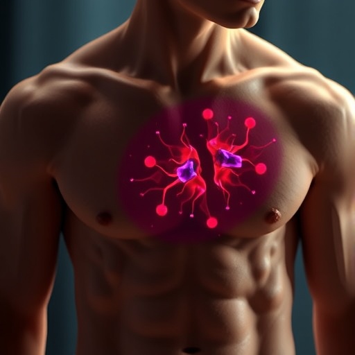
Most notable were findings pertaining to the central nervous system, where persistent post-viral effects diverged profoundly. Despite the absence of viral RNA in brain tissues from either infection, mice recovering from COVID-19 exhibited sustained neuroinflammation coupled with microvascular hemorrhages—pathological hallmarks absent in influenza survivors. This supports clinical observations of neurological dysfunction following COVID-19.
Gene expression profiling of brain tissue following SARS-CoV-2 infection revealed ongoing activation of inflammatory signaling cascades, coupled with disruption of neurotransmitter regulation pathways involving serotonin and dopamine systems. Dysregulation of these neurochemical pathways is intimately linked with the cognitive, affective, and fatigue-related symptoms emblematic of long COVID, including brain fog and mood disturbances.
The distinct neuroinflammatory signature and vasculopathy underscore the capacity of SARS-CoV-2 to elicit complex cerebrovascular immune responses, setting it apart from other respiratory viruses. This may explain the unique spectrum of neurological manifestations reported by long-haulers, advancing the paradigm of COVID-19 as a multi-system disorder with insidious intraparenchymal brain effects.
Lead author Dr. Xuebin Qin emphasized the critical need to differentiate post-viral sequelae stemming from shared respiratory insults versus those unequivocally linked to SARS-CoV-2 infection. “Our findings illustrate that although lung injury from severe respiratory viruses shares common features, the long-term cerebral consequences of COVID-19 are distinct and profound,” Qin remarked.
The study’s implications extend to the pathophysiology of vascular injury observed in COVID-19. Microbleeds and endothelial dysfunction in the brain may disrupt the blood-brain barrier integrity, facilitating a protracted inflammatory milieu. This vascular component aligns with mounting evidence associating SARS-CoV-2 with endothelialitis and thrombotic complications, fuelling neurological symptom persistence.
Supported by a targeted American Heart Association grant, the research propels forward efforts to unravel cardiovascular and cerebrovascular dimensions of post-COVID syndrome. By delineating discrete pathological trajectories in lung and brain tissue, the study lays foundational groundwork for biomarker development and therapeutics tailored to mitigate organ-specific damage.
Clinically, these mechanistic insights underscore the urgency of vigilant long-term monitoring for COVID-19 patients, even those with initially mild disease phenotypes. Understanding the divergence in repair dynamics and neuroimmune responses opens avenues for interventions aimed at enhancing lung regeneration or blunting cerebral inflammation, thus alleviating the chronic burden endured by some survivors.
The elucidation of viral interference with endogenous repair and inflammatory resolution pathways signals the need for precision medicine approaches. Future therapeutics may harness or mimic signaling that promotes airway epithelial regeneration post-infection or target neuroinflammatory mediators to forestall cognitive decline and mood disorders associated with long COVID.
As the global community continues to grapple with the enduring footprint of the COVID-19 pandemic, studies such as this provide critical molecular and cellular insights requisite for developing evidence-based strategies to address long-haul syndromes comprehensively. This knowledge is pivotal not only for improving patient outcomes but also for mitigating public health impacts stemming from prolonged morbidity.
This pioneering work propels forward the scientific dialogue, highlighting how SARS-CoV-2 uniquely disrupts neurological homeostasis and lung repair mechanisms compared to influenza, a longstanding respiratory pathogen. It lays a vital scientific milestone in decoding the complex interplay between viral infection, immune response, and tissue-specific pathology governing long-term recovery trajectories.
By integrating immunological, molecular, and histopathological analyses, the research delivers a multidimensional perspective essential for decoding the intricate consequences of post-viral syndromes. It advances the field toward precise characterization, monitoring, and ultimately targeted treatment of the lingering effects of COVID-19, charting a path toward improved health restoration for affected populations.
Subject of Research: Animals
Article Title: Characterization of Subchronic Lung and Brain Consequences Caused by Mouse-Adapted SARS-CoV-2 and Influenza A Infection of C57BL6 mice
News Publication Date: 24-Feb-2026
Web References: https://www.frontiersin.org/journals/immunology/articles/10.3389/fimmu.2026.1755141
References: Tulane University research supported by American Heart Association Long COVID Impact Project (AHA962950), National Institutes of Health grants P51OD011104-62, R01HL165265
Keywords: Long Covid, Viral infections, COVID 19, Influenza, Infectious diseases, Diseases and disorders, Health and medicine
Tags: differences between COVID-19 and influenza brain pathologyepithelial progenitor cell activation after influenzafibrotic remodeling of lung tissue post-infectionimmune activation in post-viral lungsinfluenza-induced lung injury mechanismslong-term brain impacts of COVID-19murine models in viral pathogenesis researchpost-infectious neuroinflammationprolonged respiratory symptoms after COVID-19pulmonary fibrosis after viral infectionSARS-CoV-2 neuropathological effectssubchronic effects of respiratory viruses