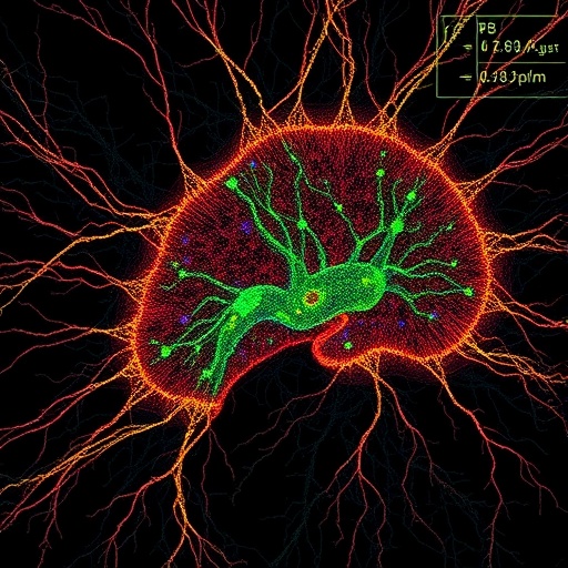
In a groundbreaking advance poised to reshape the landscape of neuroscience, researchers have unveiled a pioneering mouse brain atlas constructed through the novel lens of dendritic microenvironments. This innovative brain map transcends traditional anatomical boundaries by emphasizing the intricate spatial and functional architectures formed by dendrites—the sprawling tree-like extensions of neurons critical for synaptic integration and information processing. Published recently in Nature Neuroscience, this work offers unprecedented resolution into how neuronal circuits are organized and interconnected at the microscale, promising transformative insights into brain function, plasticity, and disease.
The creation of this atlas represents a seismic shift from classical brain mapping techniques, which primarily focus on gross cytoarchitectonic features and large-scale connectivity patterns. Unlike earlier methodologies that segmented the brain based primarily on neuron soma distribution or gross histological landmarks, this new approach capitalizes on detailed reconstructions of dendritic arborizations and their microenvironmental contexts. By doing so, the authors tap into a rich layer of structural information that mirrors the complexity and specificity of local synaptic networks, revealing how dendritic patterns define functional modules within the mammalian brain.
At the core of this research lies the sophisticated integration of high-resolution imaging modalities with advanced computational algorithms designed to decode the dense, overlapping meshwork of dendrites. Employing state-of-the-art three-dimensional microscopy combined with machine learning-driven segmentation tools, the team successfully parsed the labyrinthine structure of dendritic trees from massive imaging datasets. This enabled the generation of precise spatial distributions of dendrites across various brain regions, setting the stage for identifying microenvironmental signatures characteristic of distinct neural circuits.
One of the most striking revelations from this dendritic-based atlas is the identification of microenvironments that do not necessarily align with classical anatomical borders. These microdomains, characterized by unique dendritic density, branching complexity, and orientation patterns, suggest a finer architecture of functional compartmentalization. Such discoveries indicate that neuronal networks may be organized according to dendritic landscape principles rather than macroscopic anatomical areas alone, potentially redefining our understanding of brain region functionality.
Moreover, the dendritic microenvironments delineated in this atlas reveal nuanced layers of hierarchical organization, where local dendritic clustering correlates with specific input-output relationships and synaptic integration motifs. This finding provides a compelling structural basis for how neurons within a seemingly homogenous region can participate in diverse computations by virtue of their dendritic connectivity and spatial distribution. The atlas thereby opens a new window into dissecting cellular-level circuit mechanisms underlying sensory processing, motor control, and higher cognitive functions.
The implications of this work extend deeply into the study of neurodevelopment and neurological disorders. By mapping how dendritic microenvironments evolve during brain maturation, researchers can trace the ontogeny of functional circuits with remarkable precision. Additionally, aberrations in dendritic morphology and connectivity are central to numerous neuropathologies including autism spectrum disorders, schizophrenia, and neurodegenerative diseases. This atlas provides a critical reference framework for pinpointing microenvironmental disruptions that underpin such conditions, paving the way for targeted therapeutic interventions.
Complementing its scientific rigor, the mouse brain atlas based on dendritic microenvironments is an openly accessible resource, integrating seamlessly with existing databases and atlases. This interface empowers neuroscientists globally to superimpose dendritic organization maps with genetic, electrophysiological, and behavioral data, fostering cross-modal investigations that can unravel multifaceted brain function. The atlas thereby serves not just as a static repository but as a dynamic platform for community-driven discoveries.
The technical backbone of this endeavor encompasses several cutting-edge innovations. The imaging utilized combines volumetric fluorescence microscopy with enhanced contrast agents that selectively label dendritic structures. The authors developed custom machine learning pipelines trained on expertly annotated datasets to achieve high-fidelity dendrite segmentation despite the complexity of overlapping neurites. These tools achieved unprecedented accuracy and scalability, essential for reconstructing entire brain volumes at micrometer resolution.
Beyond structural mapping, the study also incorporates preliminary analyses linking dendritic microenvironment profiles with functional readouts obtained through in vivo imaging and electrophysiology. This multilevel approach hints at how dendritic spatial patterns influence neuronal excitability and synaptic plasticity. By correlating anatomical features with physiological data, the research underscores the integrative power of the dendritic atlas to serve as a scaffold for understanding circuit dynamics.
Further exploration of the atlas reveals striking regional variations in dendritic microarchitecture. Sensory areas such as the visual and somatosensory cortices exhibit highly stereotyped dendritic patterns supporting modality-specific computations. Conversely, association cortices and subcortical regions show more heterogeneous dendritic configurations, suggesting a structural substrate for integrative and modulatory functions. These observations set the stage for investigating how dendritic arrangements contribute to functional specialization across brain systems.
The dendritic microenvironment perspective also sheds new light on synaptic connectivity rules. Dense dendritic clustering likely facilitates local synaptic crosstalk and cooperativity, which are critical for synaptic strengthening and network plasticity. The atlas highlights that these microdomains may serve as elemental units of circuit computation, where spatial arrangement tightly governs synaptic efficacy and neural coding strategies. This paradigm challenges researchers to rethink connectivity maps beyond neuron-centric approaches, integrating dendritic spatiality as a key determinant.
This transformative atlas comes at a pivotal moment when neuroscience is increasingly embracing multidimensional approaches to decode complex brain networks. By foregrounding dendritic microenvironments, the research offers a scalable and biologically meaningful framework to dissect neural circuits at their natural operational scale. The open dissemination of this data invites the global scientific community to harness its potential, fostering innovations in brain-machine interfaces, neuroprosthetics, and artificial intelligence inspired by genuine biological blueprints.
In sum, the mouse brain atlas predicated on dendritic microenvironments stands as a landmark achievement, delivering a richly textured map that recasts our foundational understanding of brain architecture. Its technical sophistication, methodological novelty, and broad applicability promise to catalyze breakthroughs in both basic neuroscience and translational research. As investigators delve deeper into this atlas, they stand to uncover the hidden principles governing cognitive processes, neural diversity, and brain resilience.
Researchers and enthusiasts alike are poised to benefit from this resource, which blends cutting-edge imaging and computational prowess to reveal the brain’s hidden scaffolding. Future work will likely expand this approach to other species, including human brain tissues, offering a comparative lens to understand evolutionary adaptations in dendritic architecture. Ultimately, this atlas not only charts dendrites’ spatial territories but illuminates the fundamental organizational principles of the brain’s most intricate circuits.
This work also exemplifies the power of interdisciplinary collaboration, uniting neurobiology, computer science, and imaging technology to push the frontiers of brain mapping. Such integrative science underscores the importance of developing novel conceptual and technical frameworks addressing the brain’s staggering complexity. With this dendritic atlas, a new chapter opens, inviting a re-examination of neural systems through the microenvironmental tapestry orchestrated by dendrites.
As the neuroscience community digests these findings, the potential for new hypotheses, experimental designs, and clinical applications will inevitably flourish. Dendritic microenvironments offer a fertile conceptual substrate for understanding both normal cognition and pathological states, guiding future endeavors in brain repair, cognitive enhancement, and personalized medicine. The release of this atlas heralds a new era in brain mapping that promises to yield profound insights into the cellular building blocks of thought and behavior.
Subject of Research: Mouse brain structural mapping through dendritic microenvironments.
Article Title: A mouse brain atlas based on dendritic microenvironments.
Article References:
Liu, Y., Zhao, S., Yun, Z. et al. A mouse brain atlas based on dendritic microenvironments. Nat Neurosci (2025). https://doi.org/10.1038/s41593-025-02119-6
Image Credits: AI Generated
DOI: https://doi.org/10.1038/s41593-025-02119-6
Tags: brain function mappingbrain plasticity researchcomputational algorithms in neurosciencedendritic arborization analysisdendritic microenvironmentshigh-resolution imaging techniquesmicroenvironmental contexts in neuronsmouse brain atlasneuronal circuit organizationneuroscience advancementssynaptic integrationtransformative neuroscience insights


